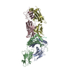
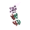
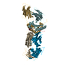
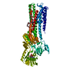
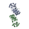
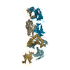
Mass: 52330.375 Da / Num. of mol.: 2 / Mutation: D325A Source method: isolated from a genetically manipulated source Source: (gene. exp.) Homo sapiens (human) / Gene: MTPAP, PAPD1 / Production host: Escherichia coli (E. coli) / Strain (production host): BL21 Rosetta (DE3) References: UniProt: Q9NVV4, polynucleotide adenylyltransferase
Has protein modification
Y
Sequence details
AMINO ACID SEQUENCE CORRESPONDING TO RESIDUES 44-538 IN UNIPROT DATABASE ENTRY Q9NVV4, PLUS A N- ...AMINO ACID SEQUENCE CORRESPONDING TO RESIDUES 44-538 IN UNIPROT DATABASE ENTRY Q9NVV4, PLUS A N-TERMINAL EXPRESSION TAG (RESIDUES 23-43), WAS USED FOR CRYSTALLIZATION. MOST OF THE RESIDUES FROM RESIDUE 134 TO 172, AND FROM RESIDUE 452 TO 490 WERE MISSING IN THE COORDINATES DUE TO LACK OF ELECTRON DENSITY. SEVERAL RESIDUES WITHIN THESE RANGES WERE MODELED AS POLY-ALA SEGMENTS, AND ARE REPRESENTED AS UNK RESIDUES 151-157, 464-468, AND 474-478 OF CHAIN A, AND UNK RESIDUES 469-478 OF CHAIN B. THESE RESIDUE NUMBERS ARE ARBITRARILY ASSIGNED. IT IS EXPECTED THAT RESIDUES 151-157 BELONG TO THE 134-172 SEGMENT, AND RESIDUES 464-468 AND 474-478 BELONG TO THE 452-490 SEGMENT.
-
Experimental details
-
Experiment
Experiment
Method: X-RAY DIFFRACTION / Number of used crystals: 1
-
Sample preparation
Crystal
Density Matthews: 2.15 Å3/Da / Density % sol: 42.8 %
Crystal grow
Temperature: 293 K / Method: microbatch Details: The precipitant solution contained 100 mM MES (pH 6.0), 6% (v/v) PEG4000, 35 mM NaCl, and 5 mM MgSO4. The protein was at 20 mg/mL concentration, and 0.5 mM MgATP was included as an additive. ...Details: The precipitant solution contained 100 mM MES (pH 6.0), 6% (v/v) PEG4000, 35 mM NaCl, and 5 mM MgSO4. The protein was at 20 mg/mL concentration, and 0.5 mM MgATP was included as an additive., microbatch, temperature 293K
In the structure databanks used in Yorodumi, some data are registered as the other names, "COVID-19 virus" and "2019-nCoV". Here are the details of the virus and the list of structure data.
Jan 31, 2019. EMDB accession codes are about to change! (news from PDBe EMDB page)
EMDB accession codes are about to change! (news from PDBe EMDB page)
The allocation of 4 digits for EMDB accession codes will soon come to an end. Whilst these codes will remain in use, new EMDB accession codes will include an additional digit and will expand incrementally as the available range of codes is exhausted. The current 4-digit format prefixed with “EMD-” (i.e. EMD-XXXX) will advance to a 5-digit format (i.e. EMD-XXXXX), and so on. It is currently estimated that the 4-digit codes will be depleted around Spring 2019, at which point the 5-digit format will come into force.
The EM Navigator/Yorodumi systems omit the EMD- prefix.
Related info.:Q: What is EMD? / ID/Accession-code notation in Yorodumi/EM Navigator
Yorodumi is a browser for structure data from EMDB, PDB, SASBDB, etc.
This page is also the successor to EM Navigator detail page, and also detail information page/front-end page for Omokage search.
The word "yorodu" (or yorozu) is an old Japanese word meaning "ten thousand". "mi" (miru) is to see.
Related info.:EMDB / PDB / SASBDB / Comparison of 3 databanks / Yorodumi Search / Aug 31, 2016. New EM Navigator & Yorodumi / Yorodumi Papers / Jmol/JSmol / Function and homology information / Changes in new EM Navigator and Yorodumi
 Movie
Movie Controller

 Yorodumi
 Open data
 Basic information
 Components
 Keywords
 Function and homology information
 Homo sapiens (human)
 X-RAY DIFFRACTION /
  Authors
 Citation
 Structure visualization
 Downloads & links
 Other downloads
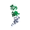

 PDBj
PDBj
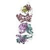
 Assembly

 Sample preparation
 / Beamline: X4A / Wavelength: 0.96863, 0.97888, 0.97926
 Processing