- PDB-3poq: Crystal structure of E.coli OmpF porin in lipidic cubic phase: sp... -
+
Open data
ID or keywords:
Loading...
-
Basic information
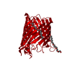
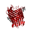
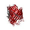
Entry
Database: PDB / ID: 3poq
Title
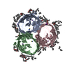
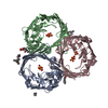
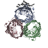
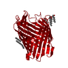
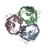
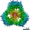
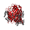
Crystal structure of E.coli OmpF porin in lipidic cubic phase: space group H32, small unit cell
Components
OmpF protein
Keywords
MEMBRANE PROTEIN / beta barrel / solute transport / pore
Function / homology
Function and homology information
colicin transmembrane transporter activity / porin activity / monoatomic ion channel complex / protein homotrimerization / pore complex / monoatomic ion channel activity / cell outer membrane / lipopolysaccharide binding / disordered domain specific binding / protein transport ...colicin transmembrane transporter activity / porin activity / monoatomic ion channel complex / protein homotrimerization / pore complex / monoatomic ion channel activity / cell outer membrane / lipopolysaccharide binding / disordered domain specific binding / protein transport / monoatomic ion transmembrane transport / lipid binding / membrane / identical protein binding Similarity search - Function
Porin, gammaproteobacterial / Porin, Gram-negative type, conserved site / General diffusion Gram-negative porins signature. / Porin domain, Gram-negative type / Gram-negative porin / Porin / Porin, Gram-negative type / : / Porin domain superfamily / Porin ...Porin, gammaproteobacterial / Porin, Gram-negative type, conserved site / General diffusion Gram-negative porins signature. / Porin domain, Gram-negative type / Gram-negative porin / Porin / Porin, Gram-negative type / : / Porin domain superfamily / Porin / Beta Barrel / Mainly Beta Similarity search - Domain/homology
(2R)-2,3-dihydroxypropyl (9Z)-octadec-9-enoate / THIOCYANATE ION / : / Outer membrane porin F Similarity search - Component
In the structure databanks used in Yorodumi, some data are registered as the other names, "COVID-19 virus" and "2019-nCoV". Here are the details of the virus and the list of structure data.
Jan 31, 2019. EMDB accession codes are about to change! (news from PDBe EMDB page)
EMDB accession codes are about to change! (news from PDBe EMDB page)
The allocation of 4 digits for EMDB accession codes will soon come to an end. Whilst these codes will remain in use, new EMDB accession codes will include an additional digit and will expand incrementally as the available range of codes is exhausted. The current 4-digit format prefixed with “EMD-” (i.e. EMD-XXXX) will advance to a 5-digit format (i.e. EMD-XXXXX), and so on. It is currently estimated that the 4-digit codes will be depleted around Spring 2019, at which point the 5-digit format will come into force.
The EM Navigator/Yorodumi systems omit the EMD- prefix.
Related info.:Q: What is EMD? / ID/Accession-code notation in Yorodumi/EM Navigator
Yorodumi is a browser for structure data from EMDB, PDB, SASBDB, etc.
This page is also the successor to EM Navigator detail page, and also detail information page/front-end page for Omokage search.
The word "yorodu" (or yorozu) is an old Japanese word meaning "ten thousand". "mi" (miru) is to see.
Related info.:EMDB / PDB / SASBDB / Comparison of 3 databanks / Yorodumi Search / Aug 31, 2016. New EM Navigator & Yorodumi / Yorodumi Papers / Jmol/JSmol / Function and homology information / Changes in new EM Navigator and Yorodumi
 Movie
Movie Controller

 Yorodumi
 Open data
 Basic information
 Components
 Keywords
 Function and homology information
 X-RAY DIFFRACTION /
  Authors
 Citation
 Structure visualization
 Downloads & links
 Other downloads


 PDBj
PDBj


 Assembly

 Mass: 356.540 Da / Num. of mol.: 23 / Source method: obtained synthetically / Formula: C21H40O4
 Mass: 58.082 Da / Num. of mol.: 3 / Source method: obtained synthetically / Formula: CNS
 Mass: 18.015 Da / Num. of mol.: 183 / Source method: isolated from a natural source / Formula: H2O
 Sample preparation
 / Beamline: X06DA / Wavelength: 1 Å
 Processing