 Movie
Movie Controller

[English] 日本語
 Yorodumi
- PDB-3plx: The crystal structure of aspartate alpha-decarboxylase from Campy... -
+ Open data

- Basic information
| Entry | Database: PDB / ID: 3plx | |||||||||
|---|---|---|---|---|---|---|---|---|---|---|
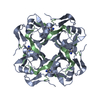
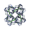
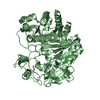
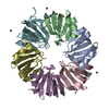
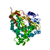
| Title | The crystal structure of aspartate alpha-decarboxylase from Campylobacter jejuni subsp. jejuni NCTC 11168 | |||||||||
 Components Components | (Aspartate 1-decarboxylase) x 2 | |||||||||
 Keywords Keywords | LYASE / STRUCTURAL GENOMICS / CENTER FOR STRUCTURAL GENOMICS OF INFECTIOUS DISEASES / CSGID / double-psi beta-barrel | |||||||||
| Function / homology |  Function and homology information Function and homology informationalanine biosynthetic process / aspartate 1-decarboxylase / aspartate 1-decarboxylase activity / pantothenate biosynthetic process / cytosol Similarity search - Function | |||||||||
| Biological species |  Campylobacter jejuni subsp. jejuni (Campylobacter) Campylobacter jejuni subsp. jejuni (Campylobacter) | |||||||||
| Method |  X-RAY DIFFRACTION / SYNCHROTRON / MOLECULAR REPLACEMENT / Resolution: 1.747 Å X-RAY DIFFRACTION / SYNCHROTRON / MOLECULAR REPLACEMENT / Resolution: 1.747 Å | |||||||||
 Authors Authors | Tan, K. / Gu, M. / Peterson, S. / Anderson, W.F. / Joachimiak, A. / Center for Structural Genomics of Infectious Diseases (CSGID) | |||||||||
 Citation Citation | Journal: To be Published Title: The crystal structure of aspartate alpha-decarboxylase from Campylobacter jejuni subsp. jejuni NCTC 11168 Authors: Tan, K. / Gu, M. / Peterson, S. / Anderson, W.F. / Joachimiak, A. | |||||||||
| History |
|
- Structure visualization
| Structure viewer | Molecule: MolmilJmol/JSmol |
|---|
- Downloads & links
-Download
| PDBx/mmCIF format | 3plx.cif.gz | 65.8 KB | Display | PDBx/mmCIF format |
|---|---|---|---|---|
| PDB format | pdb3plx.ent.gz | 48 KB | Display | PDB format |
| PDBx/mmJSON format | 3plx.json.gz | Tree view | PDBx/mmJSON format | |
| Others |  Other downloads Other downloads |
-Validation report
| Summary document | 3plx_validation.pdf.gz | 452.3 KB | Display | wwPDB validaton report |
|---|---|---|---|---|
| Full document | 3plx_full_validation.pdf.gz | 452.1 KB | Display | |
| Data in XML | 3plx_validation.xml.gz | 8.3 KB | Display | |
| Data in CIF | 3plx_validation.cif.gz | 10.7 KB | Display | |
| Arichive directory | https://data.pdbj.org/pub/pdb/validation_reports/pl/3plxftp://data.pdbj.org/pub/pdb/validation_reports/pl/3plx | HTTPS FTP |
-Related structure data
| Related structure data |  2c45S S: Starting model for refinement |
|---|---|
| Similar structure data | |
| Other databases |


-Links
 PDBj
PDBj


- Assembly
| Deposited unit | 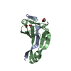
| ||||||||
|---|---|---|---|---|---|---|---|---|---|
| 1 | 
| ||||||||
| Unit cell |
|
-Components
| #1: Protein/peptide | Mass: 3007.490 Da / Num. of mol.: 1 / Fragment: UNP residues 1-24 Source method: isolated from a genetically manipulated source Source: (gene. exp.) Campylobacter jejuni subsp. jejuni (Campylobacter)Strain: NCTC 11168 / Gene: Cj0296c, panD / Plasmid: pMCSG7 / Production host: |
|---|---|
| #2: Protein | Mass: 11255.597 Da / Num. of mol.: 1 / Fragment: UNP residues 25-126 Source method: isolated from a genetically manipulated source Source: (gene. exp.) Campylobacter jejuni subsp. jejuni (Campylobacter)Strain: NCTC 11168 / Gene: Cj0296c, panD / Plasmid: pMCSG7 / Production host: |
| #3: Chemical | ChemComp-ACT /   Mass: 59.044 Da / Num. of mol.: 1 / Source method: obtained synthetically / Formula: C2H3O2 Mass: 59.044 Da / Num. of mol.: 1 / Source method: obtained synthetically / Formula: C2H3O2 |
| #4: Chemical | ChemComp-PEG /   Mass: 106.120 Da / Num. of mol.: 1 / Source method: obtained synthetically / Formula: C4H10O3 Mass: 106.120 Da / Num. of mol.: 1 / Source method: obtained synthetically / Formula: C4H10O3 |
| #5: Water | ChemComp-HOH /  Mass: 18.015 Da / Num. of mol.: 99 / Source method: isolated from a natural source / Formula: H2O Mass: 18.015 Da / Num. of mol.: 99 / Source method: isolated from a natural source / Formula: H2O |
| Compound details | THE PROTEIN IS SELF-PROCESSED AT GLY24-SER25. THE SER25 IS CONVERTED INTO A PYRUVOYL GROUP. |
| Has protein modification | Y |
| Sequence details | THE PROTEIN IS SELF-PROCESSED AT GLY24-SER25. THE SER25 IS CONVERTED INTO A PYRUVOYL GROUP. |
-Experimental details
-Experiment
| Experiment | Method: X-RAY DIFFRACTION / Number of used crystals: 1 |
|---|
- Sample preparation
| Crystal | Density Matthews: 2.82 Å3/Da / Density % sol: 56.34 % |
|---|---|
| Crystal grow | Temperature: 289 K / Method: vapor diffusion, sitting drop / pH: 7.5 Details: 0.2M Sodium Acetate, 0.1M HEPES:NaOH, 20% (W/V) PEG 3000, pH 7.5, VAPOR DIFFUSION, SITTING DROP, temperature 289K |
-Data collection
| Diffraction | Mean temperature: 100 K |
|---|---|
| Diffraction source | Source: SYNCHROTRON / Site: APS  / Beamline: 19-ID / Wavelength: 0.97921 Å / Beamline: 19-ID / Wavelength: 0.97921 Å |
| Detector | Type: ADSC QUANTUM 315r / Detector: CCD / Date: Nov 10, 2010 / Details: mirror |
| Radiation | Monochromator: Si 111 crystal / Protocol: SINGLE WAVELENGTH / Monochromatic (M) / Laue (L): M / Scattering type: x-ray |
| Radiation wavelength | Wavelength: 0.97921 Å / Relative weight: 1 |
| Reflection | Resolution: 1.75→32 Å / Num. all: 16734 / Num. obs: 16734 / % possible obs: 99.2 % / Observed criterion σ(F): 0 / Observed criterion σ(I): 0 / Redundancy: 11.5 % / Rmerge(I) obs: 0.083 / Net I/σ(I): 41.6 |
| Reflection shell | Resolution: 1.75→1.78 Å / Redundancy: 11.7 % / Rmerge(I) obs: 0.634 / Mean I/σ(I) obs: 4.97 / % possible all: 100 |
- Processing
| Software |
| |||||||||||||||||||||||||||||||||||||||||||||||||
|---|---|---|---|---|---|---|---|---|---|---|---|---|---|---|---|---|---|---|---|---|---|---|---|---|---|---|---|---|---|---|---|---|---|---|---|---|---|---|---|---|---|---|---|---|---|---|---|---|---|---|
| Refinement | Method to determine structure: MOLECULAR REPLACEMENT Starting model: PDB ENTRY 2C45 Resolution: 1.747→31.111 Å / SU ML: 0.17 / σ(F): 0.16 / Phase error: 14.08 / Stereochemistry target values: ML
| |||||||||||||||||||||||||||||||||||||||||||||||||
| Solvent computation | Shrinkage radii: 0.9 Å / VDW probe radii: 1.11 Å / Solvent model: FLAT BULK SOLVENT MODEL / Bsol: 37.132 Å2 / ksol: 0.325 e/Å3 | |||||||||||||||||||||||||||||||||||||||||||||||||
| Displacement parameters |
| |||||||||||||||||||||||||||||||||||||||||||||||||
| Refinement step | Cycle: LAST / Resolution: 1.747→31.111 Å
| |||||||||||||||||||||||||||||||||||||||||||||||||
| Refine LS restraints |
| |||||||||||||||||||||||||||||||||||||||||||||||||
| LS refinement shell |
| |||||||||||||||||||||||||||||||||||||||||||||||||
| Refinement TLS params. | Method: refined / Origin x: 8.4271 Å / Origin y: 13.6452 Å / Origin z: 29.8093 Å
| |||||||||||||||||||||||||||||||||||||||||||||||||
| Refinement TLS group | Selection details: chain A or chain B |
