 Movie
Movie Controller

[English] 日本語
 Yorodumi
- PDB-1uhe: Crystal structure of aspartate decarboxylase, isoaspargine complex -
+ Open data

- Basic information
| Entry | Database: PDB / ID: 1uhe | ||||||
|---|---|---|---|---|---|---|---|
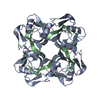
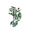
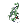
| Title | Crystal structure of aspartate decarboxylase, isoaspargine complex | ||||||
 Components Components |
| ||||||
 Keywords Keywords | LYASE / double-psi beta barrel | ||||||
| Function / homology |  Function and homology information Function and homology informationalanine biosynthetic process / aspartate 1-decarboxylase / aspartate 1-decarboxylase activity / pantothenate biosynthetic process / cytosol Similarity search - Function | ||||||
| Biological species |   Helicobacter pylori (bacteria) Helicobacter pylori (bacteria) | ||||||
| Method |  X-RAY DIFFRACTION / SYNCHROTRON / MOLECULAR REPLACEMENT / Resolution: 1.55 Å X-RAY DIFFRACTION / SYNCHROTRON / MOLECULAR REPLACEMENT / Resolution: 1.55 Å | ||||||
 Authors Authors | Lee, B.I. / Kwon, A.-R. / Han, B.W. / Suh, S.W. | ||||||
 Citation Citation | Journal: J.Mol.Biol. / Year: 2004 Title: Crystal structure of the schiff base intermediate prior to decarboxylation in the catalytic cycle of aspartate alpha-decarboxylase Authors: Lee, B.I. / Suh, S.W. | ||||||
| History |
|
- Structure visualization
| Structure viewer | Molecule: MolmilJmol/JSmol |
|---|
- Downloads & links
-Download
| PDBx/mmCIF format | 1uhe.cif.gz | 38.9 KB | Display | PDBx/mmCIF format |
|---|---|---|---|---|
| PDB format | pdb1uhe.ent.gz | 26.4 KB | Display | PDB format |
| PDBx/mmJSON format | 1uhe.json.gz | Tree view | PDBx/mmJSON format | |
| Others |  Other downloads Other downloads |
-Validation report
| Arichive directory | https://data.pdbj.org/pub/pdb/validation_reports/uh/1uheftp://data.pdbj.org/pub/pdb/validation_reports/uh/1uhe | HTTPS FTP |
|---|
-Related structure data


-Links
 PDBj
PDBj


- Assembly
| Deposited unit | 
| ||||||||
|---|---|---|---|---|---|---|---|---|---|
| 1 | 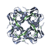
| ||||||||
| 2 | x 8
| ||||||||
| Unit cell |
| ||||||||
| Details | The biological assembly is tetramer from the monomer in the asymmetric unit by the operations: -y+1, x+1, z; -x, -y+2, z; y-1, -x+1,z |
-Components
| #1: Protein/peptide | Mass: 2806.241 Da / Num. of mol.: 1 Source method: isolated from a genetically manipulated source Source: (gene. exp.) Helicobacter pylori (bacteria) / Gene: PAND / Plasmid: pET21a / Production host: |
|---|---|
| #2: Protein | Mass: 10737.368 Da / Num. of mol.: 1 Source method: isolated from a genetically manipulated source Source: (gene. exp.) Helicobacter pylori (bacteria) / Gene: PAND / Plasmid: pET21a / Production host: |
| #3: Chemical | ChemComp-NSN /   Mass: 201.180 Da / Num. of mol.: 1 / Source method: obtained synthetically / Formula: C7H11N3O4 Mass: 201.180 Da / Num. of mol.: 1 / Source method: obtained synthetically / Formula: C7H11N3O4 |
| #4: Water | ChemComp-HOH /  Mass: 18.015 Da / Num. of mol.: 83 / Source method: isolated from a natural source / Formula: H2O Mass: 18.015 Da / Num. of mol.: 83 / Source method: isolated from a natural source / Formula: H2O |
| Has protein modification | Y |
-Experimental details
-Experiment
| Experiment | Method: X-RAY DIFFRACTION / Number of used crystals: 1 |
|---|
- Sample preparation
| Crystal | Density Matthews: 2.68 Å3/Da / Density % sol: 53.83 % |
|---|---|
| Crystal grow | Temperature: 297 K / Method: vapor diffusion, hanging drop / pH: 7.5 Details: sodium formate, pH 7.5, VAPOR DIFFUSION, HANGING DROP, temperature 297K |
-Data collection
| Diffraction | Mean temperature: 100 K |
|---|---|
| Diffraction source | Source: SYNCHROTRON / Site: PAL/PLS  / Beamline: 6B / Wavelength: 0.9795 Å / Beamline: 6B / Wavelength: 0.9795 Å |
| Detector | Type: MACSCIENCE / Detector: IMAGE PLATE |
| Radiation | Protocol: SINGLE WAVELENGTH / Monochromatic (M) / Laue (L): M / Scattering type: x-ray |
| Radiation wavelength | Wavelength: 0.9795 Å / Relative weight: 1 |
| Reflection | Resolution: 1.55→30 Å / Num. obs: 22394 / % possible obs: 95.6 % / Observed criterion σ(F): 0 / Observed criterion σ(I): 0 / Redundancy: 10 % / Biso Wilson estimate: 20.1 Å2 / Rsym value: 0.032 |
| Reflection shell | Resolution: 1.55→1.61 Å / Redundancy: 2.9 % / Mean I/σ(I) obs: 5 / Num. unique all: 2057 / Rsym value: 0.17 / % possible all: 90 |
- Processing
| Software |
| ||||||||||||||||||||
|---|---|---|---|---|---|---|---|---|---|---|---|---|---|---|---|---|---|---|---|---|---|
| Refinement | Method to determine structure: MOLECULAR REPLACEMENT / Resolution: 1.55→22.65 Å / Rfactor Rfree error: 0.005 / Data cutoff high absF: 1679240.26 / Data cutoff low absF: 0 / Isotropic thermal model: RESTRAINED / Cross valid method: THROUGHOUT / σ(F): 0
| ||||||||||||||||||||
| Solvent computation | Solvent model: FLAT MODEL / Bsol: 64.3258 Å2 / ksol: 0.387016 e/Å3 | ||||||||||||||||||||
| Displacement parameters | Biso mean: 22.7 Å2
| ||||||||||||||||||||
| Refine analyze |
| ||||||||||||||||||||
| Refinement step | Cycle: LAST / Resolution: 1.55→22.65 Å
| ||||||||||||||||||||
| Refine LS restraints |
| ||||||||||||||||||||
| LS refinement shell | Resolution: 1.55→1.65 Å / Rfactor Rfree error: 0.014 / Total num. of bins used: 6
| ||||||||||||||||||||
| Xplor file |
|
