 Movie
Movie Controller


+ Open data

- Basic information
| Entry | Database: PDB / ID: 6p1y | |||||||||
|---|---|---|---|---|---|---|---|---|---|---|
| Title | Crystal structure of Mtb aspartate decarboxylase mutant M117I | |||||||||
 Components Components |
| |||||||||
 Keywords Keywords | LYASE / Structural Genomics / TB Structural Genomics Consortium / TBSGC / POA / drug-resistance / mutant | |||||||||
| Function / homology |  Function and homology information Function and homology informationalanine biosynthetic process / aspartate 1-decarboxylase / aspartate 1-decarboxylase activity / pantothenate biosynthetic process / peptidoglycan-based cell wall / cytosol Similarity search - Function | |||||||||
| Biological species |   Mycobacterium tuberculosis (bacteria) Mycobacterium tuberculosis (bacteria) | |||||||||
| Method |  X-RAY DIFFRACTION / SYNCHROTRON / MOLECULAR REPLACEMENT / Resolution: 2.33 Å X-RAY DIFFRACTION / SYNCHROTRON / MOLECULAR REPLACEMENT / Resolution: 2.33 Å | |||||||||
 Authors Authors | Sun, Q. / Li, X. / Sacchettini, J.C. / TB Structural Genomics Consortium (TBSGC) | |||||||||
| Funding support |  United States, 2items United States, 2items
| |||||||||
 Citation Citation | Journal: Nat Commun / Year: 2020 Title: The molecular basis of pyrazinamide activity on Mycobacterium tuberculosis PanD. Authors: Sun, Q. / Li, X. / Perez, L.M. / Shi, W. / Zhang, Y. / Sacchettini, J.C. | |||||||||
| History |
|
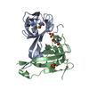
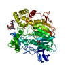
- Structure visualization
| Structure viewer | Molecule: MolmilJmol/JSmol |
|---|
- Downloads & links
-Download
| PDBx/mmCIF format | 6p1y.cif.gz | 65.2 KB | Display | PDBx/mmCIF format |
|---|---|---|---|---|
| PDB format | pdb6p1y.ent.gz | 46.1 KB | Display | PDB format |
| PDBx/mmJSON format | 6p1y.json.gz | Tree view | PDBx/mmJSON format | |
| Others |  Other downloads Other downloads |
-Validation report
| Arichive directory | https://data.pdbj.org/pub/pdb/validation_reports/p1/6p1yftp://data.pdbj.org/pub/pdb/validation_reports/p1/6p1y | HTTPS FTP |
|---|
-Related structure data
| Related structure data |  6oyyC  6oz8C  6p02C  2c45S S: Starting model for refinement C: citing same article ( |
|---|---|
| Similar structure data |


-Links
 PDBj
PDBj


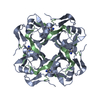
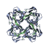
- Assembly
| Deposited unit | 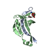
| ||||||||
|---|---|---|---|---|---|---|---|---|---|
| 1 | 
| ||||||||
| Unit cell |
|
-Components
| #1: Protein/peptide | Mass: 2751.341 Da / Num. of mol.: 1 Source method: isolated from a genetically manipulated source Source: (gene. exp.) Mycobacterium tuberculosis (strain ATCC 25618 / H37Rv) (bacteria)Strain: ATCC 25618 / H37Rv / Gene: panD, Rv3601c, MTCY07H7B.21 / Production host: |
|---|---|
| #2: Protein | Mass: 13203.739 Da / Num. of mol.: 1 / Mutation: M117I Source method: isolated from a genetically manipulated source Source: (gene. exp.) Mycobacterium tuberculosis (strain ATCC 25618 / H37Rv) (bacteria)Strain: ATCC 25618 / H37Rv / Gene: panD, Rv3601c, MTCY07H7B.21 / Production host: |
| #3: Chemical | ChemComp-NH4 /   Mass: 18.038 Da / Num. of mol.: 1 / Source method: obtained synthetically / Formula: H4N Mass: 18.038 Da / Num. of mol.: 1 / Source method: obtained synthetically / Formula: H4N |
| #4: Chemical | ChemComp-TLA /   Mass: 150.087 Da / Num. of mol.: 1 / Source method: isolated from a natural source / Formula: C4H6O6 Mass: 150.087 Da / Num. of mol.: 1 / Source method: isolated from a natural source / Formula: C4H6O6 |
| #5: Water | ChemComp-HOH /  Mass: 18.015 Da / Num. of mol.: 39 / Source method: isolated from a natural source / Formula: H2O Mass: 18.015 Da / Num. of mol.: 39 / Source method: isolated from a natural source / Formula: H2O |
| Has protein modification | Y |
-Experimental details
-Experiment
| Experiment | Method: X-RAY DIFFRACTION / Number of used crystals: 1 |
|---|
- Sample preparation
| Crystal | Density Matthews: 2.29 Å3/Da / Density % sol: 46.22 % |
|---|---|
| Crystal grow | Temperature: 290 K / Method: vapor diffusion, hanging drop / pH: 6.6 / Details: Ammonium tartrate dibasic, PEG 3350 / PH range: 6.5-7 |
-Data collection
| Diffraction | Mean temperature: 130 K / Serial crystal experiment: N |
|---|---|
| Diffraction source | Source: SYNCHROTRON / Site: APS / Beamline: 19-ID / Wavelength: 0.979 Å |
| Detector | Type: ADSC QUANTUM 315r / Detector: CCD / Date: Aug 19, 2015 |
| Radiation | Protocol: SINGLE WAVELENGTH / Monochromatic (M) / Laue (L): M / Scattering type: x-ray |
| Radiation wavelength | Wavelength: 0.979 Å / Relative weight: 1 |
| Reflection | Resolution: 2.33→36.51 Å / Num. obs: 6635 / % possible obs: 99.58 % / Redundancy: 11.3 % / Net I/σ(I): 18.88 |
| Reflection shell | Resolution: 2.33→2.414 Å / Mean I/σ(I) obs: 3.75 / Num. unique obs: 636 / % possible all: 96.95 |
- Processing
| Software |
| ||||||||||||||||||||||||||||||||||||||||
|---|---|---|---|---|---|---|---|---|---|---|---|---|---|---|---|---|---|---|---|---|---|---|---|---|---|---|---|---|---|---|---|---|---|---|---|---|---|---|---|---|---|
| Refinement | Method to determine structure: MOLECULAR REPLACEMENT Starting model: 2C45 Resolution: 2.33→36.51 Å / SU ML: 0.21 / Cross valid method: THROUGHOUT / σ(F): 1.37 / Phase error: 20.78
| ||||||||||||||||||||||||||||||||||||||||
| Solvent computation | Shrinkage radii: 0.9 Å / VDW probe radii: 1.11 Å | ||||||||||||||||||||||||||||||||||||||||
| Displacement parameters | Biso max: 136.6 Å2 / Biso mean: 38.8028 Å2 / Biso min: 20.82 Å2 | ||||||||||||||||||||||||||||||||||||||||
| Refinement step | Cycle: final / Resolution: 2.33→36.51 Å
| ||||||||||||||||||||||||||||||||||||||||
| LS refinement shell | Refine-ID: X-RAY DIFFRACTION / Rfactor Rfree error: 0
| ||||||||||||||||||||||||||||||||||||||||
| Refinement TLS params. | Method: refined / Origin x: -27.8584 Å / Origin y: -22.3682 Å / Origin z: -18.0447 Å
| ||||||||||||||||||||||||||||||||||||||||
| Refinement TLS group |
|
