: / : / : / ribonucleotide binding / DNA ligase activity / DNA ligase (ATP) / DNA ligase (ATP) activity / single-stranded DNA 3'-5' DNA exonuclease activity / DNA replication, synthesis of primer / double-strand break repair via nonhomologous end joining ...: / : / : / ribonucleotide binding / DNA ligase activity / DNA ligase (ATP) / DNA ligase (ATP) activity / single-stranded DNA 3'-5' DNA exonuclease activity / DNA replication, synthesis of primer / double-strand break repair via nonhomologous end joining / DNA-directed RNA polymerase activity / manganese ion binding / double-strand break repair / DNA recombination / DNA-directed DNA polymerase activity / nucleotide binding / magnesium ion binding / DNA binding / ATP binding / metal ion binding / plasma membrane Similarity search - Function
Alpha-Beta Plaits - #3300 / LigD polymerase domain, MtLigD-type / DNA ligase D, ligase domain / DNA ligase D, polymerase domain / : / LigD, primase-polymerase domain / DNA ligase D, 3'-phosphoesterase domain / DNA Ligase D 3'-phosphoesterase domain / DNA ligase, ATP-dependent, C-terminal / ATP dependent DNA ligase C terminal region ...Alpha-Beta Plaits - #3300 / LigD polymerase domain, MtLigD-type / DNA ligase D, ligase domain / DNA ligase D, polymerase domain / : / LigD, primase-polymerase domain / DNA ligase D, 3'-phosphoesterase domain / DNA Ligase D 3'-phosphoesterase domain / DNA ligase, ATP-dependent, C-terminal / ATP dependent DNA ligase C terminal region / ATP-dependent DNA ligase family profile. / DNA ligase, ATP-dependent, central / ATP dependent DNA ligase domain / Alpha-Beta Plaits / Nucleic acid-binding, OB-fold / 2-Layer Sandwich / Alpha Beta Similarity search - Domain/homology
: / URIDINE 5'-TRIPHOSPHATE / DNA / DNA (> 10) / Multifunctional non-homologous end joining DNA repair protein LigD / Multifunctional non-homologous end joining DNA repair protein LigD Similarity search - Component
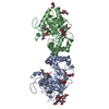
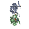
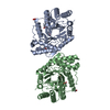
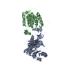
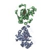
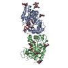
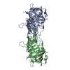
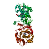
A: Putative DNA ligase-like protein B: Putative DNA ligase-like protein C: DNA 5'-D(P*GP*CP*GP*GP*C)-3' D: DNA 5'-D(*G*CP*CP*GP*CP*AP*AP*CP*GP*CP*AP*CP*G)-3' hetero molecules
In the structure databanks used in Yorodumi, some data are registered as the other names, "COVID-19 virus" and "2019-nCoV". Here are the details of the virus and the list of structure data.
Jan 31, 2019. EMDB accession codes are about to change! (news from PDBe EMDB page)
EMDB accession codes are about to change! (news from PDBe EMDB page)
The allocation of 4 digits for EMDB accession codes will soon come to an end. Whilst these codes will remain in use, new EMDB accession codes will include an additional digit and will expand incrementally as the available range of codes is exhausted. The current 4-digit format prefixed with “EMD-” (i.e. EMD-XXXX) will advance to a 5-digit format (i.e. EMD-XXXXX), and so on. It is currently estimated that the 4-digit codes will be depleted around Spring 2019, at which point the 5-digit format will come into force.
The EM Navigator/Yorodumi systems omit the EMD- prefix.
Related info.:Q: What is EMD? / ID/Accession-code notation in Yorodumi/EM Navigator
Yorodumi is a browser for structure data from EMDB, PDB, SASBDB, etc.
This page is also the successor to EM Navigator detail page, and also detail information page/front-end page for Omokage search.
The word "yorodu" (or yorozu) is an old Japanese word meaning "ten thousand". "mi" (miru) is to see.
Related info.:EMDB / PDB / SASBDB / Comparison of 3 databanks / Yorodumi Search / Aug 31, 2016. New EM Navigator & Yorodumi / Yorodumi Papers / Jmol/JSmol / Function and homology information / Changes in new EM Navigator and Yorodumi
 Movie
Movie Controller

 Yorodumi
 Open data
 Basic information
 Components
 Keywords
 Function and homology information
 Mycobacterium tuberculosis (bacteria)
 X-RAY DIFFRACTION /
  Authors
 Citation
 Structure visualization
 Downloads & links
 Other downloads
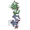

 PDBj
PDBj


 Assembly
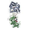


 Mass: 484.141 Da / Num. of mol.: 2 / Source method: obtained synthetically / Formula: C9H15N2O15P3 / Comment: UTP*YM
 Mass: 54.938 Da / Num. of mol.: 4 / Source method: obtained synthetically / Formula: Mn
 Sample preparation
 / Beamline: BM16 / Wavelength: 0.9787 Å
 Processing