 Movie
Movie Controller

[English] 日本語
 Yorodumi
- PDB-1vbu: Crystal structure of native xylanase 10B from Thermotoga maritima -
+ Open data

- Basic information
| Entry | Database: PDB / ID: 1vbu | ||||||
|---|---|---|---|---|---|---|---|
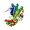
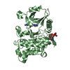
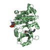
| Title | Crystal structure of native xylanase 10B from Thermotoga maritima | ||||||
 Components Components | endo-1,4-beta-xylanase B | ||||||
 Keywords Keywords | HYDROLASE / xylanase 10B | ||||||
| Function / homology |  Function and homology information Function and homology informationendo-1,4-beta-xylanase / endo-1,4-beta-xylanase activity / xylan catabolic process Similarity search - Function | ||||||
| Biological species |   Thermotoga maritima (bacteria) Thermotoga maritima (bacteria) | ||||||
| Method |  X-RAY DIFFRACTION / SYNCHROTRON / MOLECULAR REPLACEMENT / Resolution: 1.8 Å X-RAY DIFFRACTION / SYNCHROTRON / MOLECULAR REPLACEMENT / Resolution: 1.8 Å | ||||||
 Authors Authors | Ihsanawati / Kumasaka, T. / Kaneko, T. / Nakamura, S. / Tanaka, N. | ||||||
 Citation Citation | Journal: Proteins / Year: 2005 Title: Structural basis of the substrate subsite and the highly thermal stability of xylanase 10B from Thermotoga maritima MSB8 Authors: Ihsanawati / Kumasaka, T. / Kaneko, T. / Morokuma, C. / Yatsunami, R. / Sato, T. / Nakamura, S. / Tanaka, N. #1: Journal: ACTA CRYSTALLOGR.,SECT.D / Year: 2003 Title: Crystallization and preliminary X-ray studies of xylanse 10B from Thermotoga maritima Authors: Ihsanawati / Kumasaka, T. / Kaneko, T. / Morokuma, C. / Nakamura, S. / Tanaka, N. | ||||||
| History |
|
- Structure visualization
| Structure viewer | Molecule: MolmilJmol/JSmol |
|---|
- Downloads & links
-Download
| PDBx/mmCIF format | 1vbu.cif.gz | 156.9 KB | Display | PDBx/mmCIF format |
|---|---|---|---|---|
| PDB format | pdb1vbu.ent.gz | 122.1 KB | Display | PDB format |
| PDBx/mmJSON format | 1vbu.json.gz | Tree view | PDBx/mmJSON format | |
| Others |  Other downloads Other downloads |
-Validation report
| Arichive directory | https://data.pdbj.org/pub/pdb/validation_reports/vb/1vbuftp://data.pdbj.org/pub/pdb/validation_reports/vb/1vbu | HTTPS FTP |
|---|
-Related structure data


-Links
 PDBj
PDBj


- Assembly
| Deposited unit | 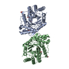
| ||||||||
|---|---|---|---|---|---|---|---|---|---|
| 1 | 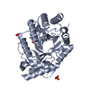
| ||||||||
| 2 | 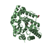
| ||||||||
| Unit cell |
|
-Components
| #1: Protein | Mass: 38700.852 Da / Num. of mol.: 2 Source method: isolated from a genetically manipulated source Source: (gene. exp.) Thermotoga maritima (bacteria) / Plasmid: pET21b / Production host: #2: Chemical | ChemComp-SO4 / |   Mass: 96.063 Da / Num. of mol.: 1 / Source method: obtained synthetically / Formula: SO4 Mass: 96.063 Da / Num. of mol.: 1 / Source method: obtained synthetically / Formula: SO4#3: Chemical | ChemComp-ACY /   Mass: 60.052 Da / Num. of mol.: 6 / Source method: obtained synthetically / Formula: C2H4O2 Mass: 60.052 Da / Num. of mol.: 6 / Source method: obtained synthetically / Formula: C2H4O2#4: Chemical |   Mass: 92.094 Da / Num. of mol.: 2 / Source method: obtained synthetically / Formula: C3H8O3 Mass: 92.094 Da / Num. of mol.: 2 / Source method: obtained synthetically / Formula: C3H8O3#5: Water | ChemComp-HOH / |  Mass: 18.015 Da / Num. of mol.: 618 / Source method: isolated from a natural source / Formula: H2O Mass: 18.015 Da / Num. of mol.: 618 / Source method: isolated from a natural source / Formula: H2O |
|---|
-Experimental details
-Experiment
| Experiment | Method: X-RAY DIFFRACTION / Number of used crystals: 1 |
|---|
- Sample preparation
| Crystal | Density Matthews: 2.2 Å3/Da / Density % sol: 43.54 % |
|---|---|
| Crystal grow | Temperature: 293 K / Method: vapor diffusion, hanging drop / pH: 4.3 Details: PEG 8000, sodium acetate, lithium sulfate, pH 4.3, VAPOR DIFFUSION, HANGING DROP, temperature 293K |
-Data collection
| Diffraction | Mean temperature: 100 K |
|---|---|
| Diffraction source | Source: SYNCHROTRON / Site: SPring-8  / Beamline: BL38B1 / Wavelength: 1 Å / Beamline: BL38B1 / Wavelength: 1 Å |
| Detector | Type: ADSC QUANTUM 4 / Detector: CCD / Date: Mar 25, 2003 |
| Radiation | Monochromator: graphite / Protocol: SINGLE WAVELENGTH / Monochromatic (M) / Laue (L): M / Scattering type: x-ray |
| Radiation wavelength | Wavelength: 1 Å / Relative weight: 1 |
| Reflection | Resolution: 1.8→57 Å / Num. all: 64816 / Num. obs: 64816 / % possible obs: 100 % / Observed criterion σ(F): 0 / Observed criterion σ(I): 0 / Biso Wilson estimate: 13.1 Å2 |
| Reflection shell | Resolution: 1.8→1.87 Å / % possible all: 96.8 |
- Processing
| Software |
| |||||||||||||||||||||||||
|---|---|---|---|---|---|---|---|---|---|---|---|---|---|---|---|---|---|---|---|---|---|---|---|---|---|---|
| Refinement | Method to determine structure: MOLECULAR REPLACEMENT / Resolution: 1.8→29.53 Å / Rfactor Rfree error: 0.004 / Data cutoff high absF: 1006234.16 / Data cutoff low absF: 0 / Isotropic thermal model: RESTRAINED / Cross valid method: THROUGHOUT / σ(F): 0 / Stereochemistry target values: Engh & Huber
| |||||||||||||||||||||||||
| Solvent computation | Solvent model: FLAT MODEL / Bsol: 49.1769 Å2 / ksol: 0.376806 e/Å3 | |||||||||||||||||||||||||
| Displacement parameters | Biso mean: 14.7 Å2
| |||||||||||||||||||||||||
| Refine analyze |
| |||||||||||||||||||||||||
| Refinement step | Cycle: LAST / Resolution: 1.8→29.53 Å
| |||||||||||||||||||||||||
| Refine LS restraints |
| |||||||||||||||||||||||||
| LS refinement shell | Resolution: 1.8→1.91 Å / Rfactor Rfree error: 0.011 / Total num. of bins used: 6
| |||||||||||||||||||||||||
| Xplor file |
|
