 Movie
Movie Controller

[English] 日本語
 Yorodumi
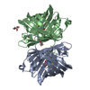
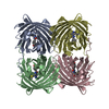
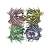
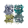
- PDB-3pj7: Crystal structure of far-red fluorescent protein Katushka crystal... -
+ Open data

- Basic information
| Entry | Database: PDB / ID: 3pj7 | |||||||||
|---|---|---|---|---|---|---|---|---|---|---|
| Title | Crystal structure of far-red fluorescent protein Katushka crystallized at pH 8.5 | |||||||||
 Components Components | Red fluorescent protein eqFP578 | |||||||||
 Keywords Keywords | FLUORESCENT PROTEIN / Katushka / far-red fluorescent protein / beta-barrel / Biomarker / Mutant variant of eqFP578 / Met-Tyr-Gly chromophore | |||||||||
| Function / homology | Green Fluorescent Protein / Green fluorescent protein / Green fluorescent protein-related / Green fluorescent protein / Green fluorescent protein / bioluminescence / Beta Barrel / Mainly Beta / Red fluorescent protein eqFP578 Function and homology information Function and homology information | |||||||||
| Biological species |  Entacmaea quadricolor (sea anemone) Entacmaea quadricolor (sea anemone) | |||||||||
| Method |  X-RAY DIFFRACTION / SYNCHROTRON / MOLECULAR REPLACEMENT / Resolution: 1.85 Å X-RAY DIFFRACTION / SYNCHROTRON / MOLECULAR REPLACEMENT / Resolution: 1.85 Å | |||||||||
 Authors Authors | Pletnev, S. / Pletneva, N.V. / Pletnev, V.Z. | |||||||||
 Citation Citation | Journal: Protein Sci. / Year: 2011 Title: Crystallographic study of red fluorescent protein eqFP578 and its far-red variant Katushka reveals opposite pH-induced isomerization of chromophore. Authors: Pletneva, N.V. / Pletnev, V.Z. / Shemiakina, I.I. / Chudakov, D.M. / Artemyev, I. / Wlodawer, A. / Dauter, Z. / Pletnev, S. | |||||||||
| History |
|
- Structure visualization
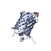
| Structure viewer | Molecule: MolmilJmol/JSmol |
|---|
- Downloads & links
-Download
| PDBx/mmCIF format | 3pj7.cif.gz | 194.9 KB | Display | PDBx/mmCIF format |
|---|---|---|---|---|
| PDB format | pdb3pj7.ent.gz | 157.1 KB | Display | PDB format |
| PDBx/mmJSON format | 3pj7.json.gz | Tree view | PDBx/mmJSON format | |
| Others |  Other downloads Other downloads |
-Validation report
| Arichive directory | https://data.pdbj.org/pub/pdb/validation_reports/pj/3pj7ftp://data.pdbj.org/pub/pdb/validation_reports/pj/3pj7 | HTTPS FTP |
|---|
-Related structure data


-Links
 PDBj
PDBj

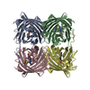
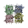
- Assembly
| Deposited unit | 
| ||||||||
|---|---|---|---|---|---|---|---|---|---|
| 1 |
| ||||||||
| Unit cell |
|
-Components
| #1: Protein | Mass: 25989.564 Da / Num. of mol.: 4 Source method: isolated from a genetically manipulated source Source: (gene. exp.) Entacmaea quadricolor (sea anemone) / Plasmid: pQE30 / Production host:  #2: Water | ChemComp-HOH / |  Mass: 18.015 Da / Num. of mol.: 462 / Source method: isolated from a natural source / Formula: H2O Mass: 18.015 Da / Num. of mol.: 462 / Source method: isolated from a natural source / Formula: H2OHas protein modification | Y | |
|---|
-Experimental details
-Experiment
| Experiment | Method: X-RAY DIFFRACTION / Number of used crystals: 1 |
|---|
- Sample preparation
| Crystal | Density Matthews: 2.33 Å3/Da / Density % sol: 47.25 % |
|---|---|
| Crystal grow | Temperature: 293 K / Method: vapor diffusion, hanging drop / pH: 8.5 Details: 24% w/v PEG4000, 0.16 M MgCl2, 0.08 M TRIS hydrochloride pH 8.5, 20% v/v glycerol, VAPOR DIFFUSION, HANGING DROP, temperature 293K |
-Data collection
| Diffraction | Mean temperature: 100 K | |||||||||||||||||||||||||||||||||||||||||||||||||||||||||||||||||||||||||||||
|---|---|---|---|---|---|---|---|---|---|---|---|---|---|---|---|---|---|---|---|---|---|---|---|---|---|---|---|---|---|---|---|---|---|---|---|---|---|---|---|---|---|---|---|---|---|---|---|---|---|---|---|---|---|---|---|---|---|---|---|---|---|---|---|---|---|---|---|---|---|---|---|---|---|---|---|---|---|---|
| Diffraction source | Source: SYNCHROTRON / Site: APS  / Beamline: 22-ID / Wavelength: 1 Å / Beamline: 22-ID / Wavelength: 1 Å | |||||||||||||||||||||||||||||||||||||||||||||||||||||||||||||||||||||||||||||
| Detector | Type: MARMOSAIC 300 mm CCD / Detector: CCD / Details: mirrors | |||||||||||||||||||||||||||||||||||||||||||||||||||||||||||||||||||||||||||||
| Radiation | Protocol: SINGLE WAVELENGTH / Monochromatic (M) / Laue (L): M / Scattering type: x-ray | |||||||||||||||||||||||||||||||||||||||||||||||||||||||||||||||||||||||||||||
| Radiation wavelength | Wavelength: 1 Å / Relative weight: 1 | |||||||||||||||||||||||||||||||||||||||||||||||||||||||||||||||||||||||||||||
| Reflection | Resolution: 1.8→30 Å / Num. obs: 88163 / % possible obs: 100 % / Redundancy: 5 % / Rmerge(I) obs: 0.096 / Χ2: 1.156 / Net I/σ(I): 8.4 | |||||||||||||||||||||||||||||||||||||||||||||||||||||||||||||||||||||||||||||
| Reflection shell |
|
- Processing
| Software |
| ||||||||||||||||||||||||||||||||||||||||||||||||||||||||||||||||||||||||||||||||||||||||||
|---|---|---|---|---|---|---|---|---|---|---|---|---|---|---|---|---|---|---|---|---|---|---|---|---|---|---|---|---|---|---|---|---|---|---|---|---|---|---|---|---|---|---|---|---|---|---|---|---|---|---|---|---|---|---|---|---|---|---|---|---|---|---|---|---|---|---|---|---|---|---|---|---|---|---|---|---|---|---|---|---|---|---|---|---|---|---|---|---|---|---|---|
| Refinement | Method to determine structure: MOLECULAR REPLACEMENT / Resolution: 1.85→24.99 Å / Cor.coef. Fo:Fc: 0.968 / Cor.coef. Fo:Fc free: 0.94 / WRfactor Rfree: 0.2551 / WRfactor Rwork: 0.1991 / Occupancy max: 1 / Occupancy min: 0.3 / FOM work R set: 0.7706 / SU B: 4.779 / SU ML: 0.137 / SU R Cruickshank DPI: 0.1467 / SU Rfree: 0.1453 / Cross valid method: THROUGHOUT / σ(F): 0 / ESU R Free: 0.148 / Stereochemistry target values: MAXIMUM LIKELIHOOD / Details: HYDROGENS HAVE BEEN ADDED IN THE RIDING POSITIONS
| ||||||||||||||||||||||||||||||||||||||||||||||||||||||||||||||||||||||||||||||||||||||||||
| Solvent computation | Ion probe radii: 0.8 Å / Shrinkage radii: 0.8 Å / VDW probe radii: 1.2 Å / Solvent model: MASK | ||||||||||||||||||||||||||||||||||||||||||||||||||||||||||||||||||||||||||||||||||||||||||
| Displacement parameters | Biso max: 69.2 Å2 / Biso mean: 34.6951 Å2 / Biso min: 10.94 Å2
| ||||||||||||||||||||||||||||||||||||||||||||||||||||||||||||||||||||||||||||||||||||||||||
| Refinement step | Cycle: LAST / Resolution: 1.85→24.99 Å
| ||||||||||||||||||||||||||||||||||||||||||||||||||||||||||||||||||||||||||||||||||||||||||
| Refine LS restraints |
| ||||||||||||||||||||||||||||||||||||||||||||||||||||||||||||||||||||||||||||||||||||||||||
| LS refinement shell | Resolution: 1.85→1.898 Å / Total num. of bins used: 20
|
