 Movie
Movie Controller

[English] 日本語
 Yorodumi
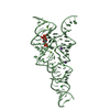
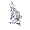
- PDB-3phs: Crystal Structure of GBS52, the minor pilin in gram-positive path... -
+ Open data

- Basic information
| Entry | Database: PDB / ID: 3phs | |||||||||
|---|---|---|---|---|---|---|---|---|---|---|
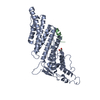
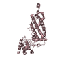
| Title | Crystal Structure of GBS52, the minor pilin in gram-positive pathogen Streptococcus agalactiae | |||||||||
 Components Components | Cell wall surface anchor family protein | |||||||||
 Keywords Keywords | Structural protein / CELL AHDESION / Ig-like fold / IgG-rev fold / GBS52 / Streptococcus agalactiae / gram-positive pilins / adhesions / CELL ADHESION / cell wall anchoring / adhesion / pilus subunit / isopeptide bond / Gram-positive bacterial cell wall | |||||||||
| Function / homology |  Function and homology information Function and homology informationPrealbumin-like fold domain / Prealbumin-like fold domain / LPXTG cell wall anchor motif / LPXTG cell wall anchor domain / Immunoglobulin-like fold / Immunoglobulins / Immunoglobulin-like / Sandwich / Mainly Beta Similarity search - Domain/homology | |||||||||
| Biological species |  Streptococcus agalactiae (bacteria) Streptococcus agalactiae (bacteria) | |||||||||
| Method |  X-RAY DIFFRACTION / MIR / Resolution: 1.8 Å X-RAY DIFFRACTION / MIR / Resolution: 1.8 Å | |||||||||
 Authors Authors | Narayana, S.V.L. / Krishnan, V. | |||||||||
 Citation Citation | Journal: Structure / Year: 2007 Title: An IgG-like domain in the minor pilin GBS52 of Streptococcus agalactiae mediates lung epithelial cell adhesion. Authors: Krishnan, V. / Gaspar, A.H. / Ye, N. / Mandlik, A. / Ton-That, H. / Narayana, S.V. | |||||||||
| History |
|
- Structure visualization
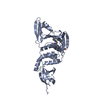
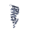
| Structure viewer | Molecule: MolmilJmol/JSmol |
|---|
- Downloads & links
-Download
| PDBx/mmCIF format | 3phs.cif.gz | 64.1 KB | Display | PDBx/mmCIF format |
|---|---|---|---|---|
| PDB format | pdb3phs.ent.gz | 46.2 KB | Display | PDB format |
| PDBx/mmJSON format | 3phs.json.gz | Tree view | PDBx/mmJSON format | |
| Others |  Other downloads Other downloads |
-Validation report
| Arichive directory | https://data.pdbj.org/pub/pdb/validation_reports/ph/3phsftp://data.pdbj.org/pub/pdb/validation_reports/ph/3phs | HTTPS FTP |
|---|
-Related structure data
| Related structure data | |
|---|---|
| Similar structure data |

-Links
 PDBj
PDBj- Assembly
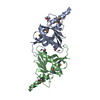
| Deposited unit | 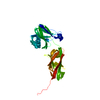
| ||||||||
|---|---|---|---|---|---|---|---|---|---|
| 1 |
| ||||||||
| Unit cell |
|
-Components
| #1: Protein | Mass: 27692.242 Da / Num. of mol.: 1 / Fragment: GBS052 (unp residues 31-267) Source method: isolated from a genetically manipulated source Source: (gene. exp.) Streptococcus agalactiae (bacteria) / Strain: 2603V/R / Gene: GBS052, SAG0646 / Plasmid: pQE30 / Production host: |
|---|---|
| #2: Water | ChemComp-HOH /  Mass: 18.015 Da / Num. of mol.: 215 / Source method: isolated from a natural source / Formula: H2O Mass: 18.015 Da / Num. of mol.: 215 / Source method: isolated from a natural source / Formula: H2O |
| Has protein modification | Y |
-Experimental details
-Experiment
| Experiment | Method: X-RAY DIFFRACTION / Number of used crystals: 1 |
|---|
- Sample preparation
| Crystal | Density Matthews: 2.34 Å3/Da / Density % sol: 47.34 % |
|---|---|
| Crystal grow | Temperature: 277 K / Method: vapor diffusion, hanging drop / pH: 7 Details: 20% PEG1500, 0.1M HEPES buffer, 0.2M Ammonium Citrate, 0.01M CaCl2, pH 7.0, VAPOR DIFFUSION, HANGING DROP, temperature 277K |
-Data collection
| Diffraction | Mean temperature: 100 K |
|---|---|
| Diffraction source | Source: ROTATING ANODE / Type: RIGAKU / Wavelength: 1.5418 Å |
| Detector | Type: RIGAKU RAXIS IV / Detector: IMAGE PLATE / Date: Jul 31, 2005 |
| Radiation | Protocol: SINGLE WAVELENGTH / Monochromatic (M) / Laue (L): M / Scattering type: x-ray |
| Radiation wavelength | Wavelength: 1.5418 Å / Relative weight: 1 |
| Reflection | Resolution: 1.8→26.81 Å / Num. all: 23655 / Num. obs: 23655 / % possible obs: 99.4 % / Redundancy: 3.01 % / Rmerge(I) obs: 0.048 / Net I/σ(I): 14.3 |
| Reflection shell | Resolution: 1.8→1.86 Å / Redundancy: 2.33 % / Rmerge(I) obs: 0.157 / Mean I/σ(I) obs: 4.6 / % possible all: 97.3 |
- Processing
| Software |
| |||||||||||||||||||||||||||||||||||||||||||||||||||||||||||||||||
|---|---|---|---|---|---|---|---|---|---|---|---|---|---|---|---|---|---|---|---|---|---|---|---|---|---|---|---|---|---|---|---|---|---|---|---|---|---|---|---|---|---|---|---|---|---|---|---|---|---|---|---|---|---|---|---|---|---|---|---|---|---|---|---|---|---|---|
| Refinement | Method to determine structure: MIR / Resolution: 1.8→26.81 Å / Cor.coef. Fo:Fc: 0.947 / Cor.coef. Fo:Fc free: 0.928 / SU B: 2.643 / SU ML: 0.083 / Cross valid method: THROUGHOUT / ESU R Free: 0.129 / Stereochemistry target values: MAXIMUM LIKELIHOOD / Details: HYDROGENS HAVE BEEN ADDED IN THE RIDING POSITIONS
| |||||||||||||||||||||||||||||||||||||||||||||||||||||||||||||||||
| Solvent computation | Ion probe radii: 0.8 Å / Shrinkage radii: 0.8 Å / VDW probe radii: 1.4 Å / Solvent model: MASK | |||||||||||||||||||||||||||||||||||||||||||||||||||||||||||||||||
| Displacement parameters | Biso mean: 18.941 Å2
| |||||||||||||||||||||||||||||||||||||||||||||||||||||||||||||||||
| Refinement step | Cycle: LAST / Resolution: 1.8→26.81 Å
| |||||||||||||||||||||||||||||||||||||||||||||||||||||||||||||||||
| Refine LS restraints |
| |||||||||||||||||||||||||||||||||||||||||||||||||||||||||||||||||
| LS refinement shell | Resolution: 1.8→1.847 Å / Total num. of bins used: 20
|
