Monochromator: SAGITALLY FOCUSED Si(111) / Protocol: SINGLE WAVELENGTH / Monochromatic (M) / Laue (L): M / Scattering type: x-ray
Radiation wavelength
Wavelength: 0.97937 Å / Relative weight: 1
Reflection
Redundancy: 23.8 % / Av σ(I) over netI: 64.13 / Number: 486484 / Rmerge(I) obs: 0.074 / Χ2: 1.59 / D res high: 1.85 Å / D res low: 50 Å / Num. obs: 20449 / % possible obs: 99.4
D res high: 1.9 Å / D res low: 50 Å / FOM : 0.3 / FOM acentric: 0.368 / FOM centric: 0 / Reflection: 19288 / Reflection acentric: 15730 / Reflection centric: 3558
Phasing MAD set
R cullis acentric: 1.82 / R cullis centric: 1 / Highest resolution: 1.9 Å / Lowest resolution: 50 Å / Loc acentric: 0.1 / Loc centric: 0.1 / Power acentric: 0 / Power centric: 0 / Reflection acentric: 15730 / Reflection centric: 3558
Phasing MAD set shell
ID: 1 / R cullis centric: 1 / Power acentric: 0 / Power centric: 0
Resolution (Å)
R cullis acentric
Loc acentric
Loc centric
Reflection acentric
Reflection centric
12.01-50
1.53
0.4
0.2
30
69
6.82-12.01
1.64
0.3
0.2
217
181
4.76-6.82
1.57
0.3
0.1
592
290
3.66-4.76
1.08
0.2
0.2
1145
393
2.97-3.66
1.34
0.1
0.1
1887
498
2.5-2.97
2.36
0.1
0
2801
613
2.16-2.5
3.01
0.1
0
3894
718
1.9-2.16
4.76
0
0
5164
796
Phasing MAD set site
Atom type symbol: Se / Occupancy iso: 0
ID
B iso
Fract x
Fract y
Fract z
Occupancy
1
37.8733
0.136
0.765
0.054
4.066
2
54.8932
0.068
0.631
-0.036
3.432
3
105.2141
0.021
0.673
-0.085
3.171
Phasing MAD shell
Resolution (Å)
FOM
FOM acentric
FOM centric
Reflection
Reflection acentric
Reflection centric
12.01-50
0.165
0.544
0
99
30
69
6.82-12.01
0.359
0.658
0
398
217
181
4.76-6.82
0.405
0.604
0
882
592
290
3.66-4.76
0.447
0.601
0
1538
1145
393
2.97-3.66
0.448
0.566
0
2385
1887
498
2.5-2.97
0.398
0.485
0
3414
2801
613
2.16-2.5
0.293
0.347
0
4612
3894
718
1.9-2.16
0.135
0.156
0
5960
5164
796
Phasing dm
Method: Solvent flattening and Histogram matching / Reflection: 20352
Phasing dm shell
Resolution (Å)
Delta phi final
FOM
Reflection
6.8-100
73.4
0.753
506
5.32-6.8
63.9
0.86
504
4.61-5.32
59.6
0.88
511
4.16-4.61
65.2
0.894
509
3.84-4.16
57.8
0.889
524
3.59-3.84
58.6
0.898
541
3.38-3.59
60.4
0.884
579
3.2-3.38
61.6
0.895
608
3.05-3.2
60.8
0.892
648
2.92-3.05
59.1
0.88
660
2.8-2.92
64.5
0.886
702
2.7-2.8
59.9
0.874
722
2.6-2.7
60.3
0.883
740
2.52-2.6
61.2
0.892
756
2.44-2.52
61.9
0.878
799
2.38-2.44
61.3
0.885
815
2.31-2.38
61.4
0.89
835
2.25-2.31
67.4
0.88
849
2.2-2.25
67
0.898
856
2.15-2.2
62
0.886
894
2.1-2.15
67.1
0.873
909
2.05-2.1
69.7
0.869
939
2.01-2.05
72
0.884
925
1.97-2.01
75.9
0.862
976
1.94-1.97
76.9
0.844
974
1.9-1.94
76.8
0.808
976
1.85-1.9
88.5
0.751
1095
-
Processing
Software
Name
Version
Classification
NB
DENZO
datareduction
SCALEPACK
datascaling
MLPHARE
phasing
DM
6.1
phasing
REFMAC
refinement
PDB_EXTRACT
3.1
dataextraction
SBC-Collect
datacollection
HKL-3000
datareduction
HKL-3000
datascaling
HKL-3000
phasing
SHELXD
phasing
SHELXE
modelbuilding
SOLVE
phasing
RESOLVE
phasing
ARP/wARP
modelbuilding
CCP4
phasing
O
modelbuilding
Coot
modelbuilding
Refinement
Method to determine structure: SAD / Resolution: 1.86→34.43 Å / Cor.coef. Fo:Fc: 0.966 / Cor.coef. Fo:Fc free: 0.961 / WRfactor Rfree: 0.1871 / WRfactor Rwork: 0.166 / Occupancy max: 1 / Occupancy min: 0.3 / FOM work R set: 0.9098 / SU B: 4.157 / SU ML: 0.057 / SU R Cruickshank DPI: 0.0999 / SU Rfree: 0.0951 / Cross valid method: THROUGHOUT / σ(F): 0 / ESU R Free: 0.095 Stereochemistry target values: MAXIMUM LIKELIHOOD WITH PHASES Details: HYDROGENS HAVE BEEN ADDED IN THE RIDING POSITIONS U VALUES : WITH TLS ADDED
Rfactor
Num. reflection
% reflection
Selection details
Rfree
0.1904
1034
5.1 %
RANDOM
Rwork
0.1703
-
-
-
all
0.1713
20287
-
-
obs
0.1713
20287
98.47 %
-
Solvent computation
Ion probe radii: 0.8 Å / Shrinkage radii: 0.8 Å / VDW probe radii: 1.4 Å / Solvent model: MASK
In the structure databanks used in Yorodumi, some data are registered as the other names, "COVID-19 virus" and "2019-nCoV". Here are the details of the virus and the list of structure data.
Jan 31, 2019. EMDB accession codes are about to change! (news from PDBe EMDB page)
EMDB accession codes are about to change! (news from PDBe EMDB page)
The allocation of 4 digits for EMDB accession codes will soon come to an end. Whilst these codes will remain in use, new EMDB accession codes will include an additional digit and will expand incrementally as the available range of codes is exhausted. The current 4-digit format prefixed with “EMD-” (i.e. EMD-XXXX) will advance to a 5-digit format (i.e. EMD-XXXXX), and so on. It is currently estimated that the 4-digit codes will be depleted around Spring 2019, at which point the 5-digit format will come into force.
The EM Navigator/Yorodumi systems omit the EMD- prefix.
Related info.:Q: What is EMD? / ID/Accession-code notation in Yorodumi/EM Navigator
Yorodumi is a browser for structure data from EMDB, PDB, SASBDB, etc.
This page is also the successor to EM Navigator detail page, and also detail information page/front-end page for Omokage search.
The word "yorodu" (or yorozu) is an old Japanese word meaning "ten thousand". "mi" (miru) is to see.
Related info.:EMDB / PDB / SASBDB / Comparison of 3 databanks / Yorodumi Search / Aug 31, 2016. New EM Navigator & Yorodumi / Yorodumi Papers / Jmol/JSmol / Function and homology information / Changes in new EM Navigator and Yorodumi
 Movie
Movie Controller

 Yorodumi
 Open data
 Basic information
 Components
 Keywords
 Function and homology information
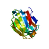
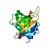
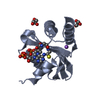
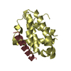
 Streptococcus thermophilus (bacteria)
 X-RAY DIFFRACTION /
  Authors
 Citation
 Structure visualization
 Downloads & links
 Other downloads


 PDBj
PDBj Assembly

 Mass: 207.290 Da / Num. of mol.: 1 / Source method: obtained synthetically / Formula: C8H17NO3S / Comment: pH buffer*YM
 Mass: 62.068 Da / Num. of mol.: 10 / Source method: obtained synthetically / Formula: C2H6O2
 Mass: 22.990 Da / Num. of mol.: 2 / Source method: obtained synthetically / Formula: Na
 Mass: 18.015 Da / Num. of mol.: 160 / Source method: isolated from a natural source / Formula: H2O
 Sample preparation
 / Beamline: 19-BM / Wavelength: 0.97937 Å
 Processing