 Movie
Movie Controller

[English] 日本語
 Yorodumi
- PDB-1fil: HUMAN PLATELET PROFILIN I CRYSTALLIZED IN HIGH SALT ACTIN-BINDING... -
+ Open data

- Basic information
| Entry | Database: PDB / ID: 1fil | ||||||
|---|---|---|---|---|---|---|---|
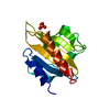
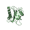
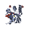
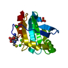
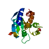
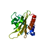
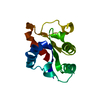
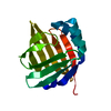
| Title | HUMAN PLATELET PROFILIN I CRYSTALLIZED IN HIGH SALT ACTIN-BINDING PROTEIN | ||||||
 Components Components | PROFILIN | ||||||
 Keywords Keywords | CONTRACTILE PROTEIN / ACETYLATION / ACTIN-BINDING PROTEIN / MULTIGENE FAMILY | ||||||
| Function / homology |  Function and homology information Function and homology informationsynapse maturation / adenyl-nucleotide exchange factor activity / negative regulation of actin filament bundle assembly / modification of postsynaptic actin cytoskeleton / positive regulation of actin filament bundle assembly / regulation of actin filament polymerization / Signaling by ROBO receptors / negative regulation of stress fiber assembly / proline-rich region binding / PCP/CE pathway ...synapse maturation / adenyl-nucleotide exchange factor activity / negative regulation of actin filament bundle assembly / modification of postsynaptic actin cytoskeleton / positive regulation of actin filament bundle assembly / regulation of actin filament polymerization / Signaling by ROBO receptors / negative regulation of stress fiber assembly / proline-rich region binding / PCP/CE pathway / positive regulation of ruffle assembly / positive regulation of actin filament polymerization / positive regulation of epithelial cell migration / actin monomer binding / phosphotyrosine residue binding / phosphatidylinositol-4,5-bisphosphate binding / neural tube closure / RHO GTPases Activate Formins / modulation of chemical synaptic transmission / small GTPase binding / Platelet degranulation / actin binding / actin cytoskeleton organization / blood microparticle / cell cortex / cytoskeleton / protein stabilization / cadherin binding / focal adhesion / regulation of transcription by RNA polymerase II / glutamatergic synapse / RNA binding / extracellular exosome / membrane / nucleus / cytoplasm / cytosol Similarity search - Function | ||||||
| Biological species |  Homo sapiens (human) Homo sapiens (human) | ||||||
| Method |  X-RAY DIFFRACTION / molecular replacement / Resolution: 2 Å X-RAY DIFFRACTION / molecular replacement / Resolution: 2 Å | ||||||
 Authors Authors | Fedorov, A.A. / Pollard, T.D. / Almo, S.C. | ||||||
 Citation Citation | Journal: To be Published Title: Crystal Structure of Human Profilin at 2.0 Angstroms Resolution Authors: Fedorov, A.A. / Pollard, T.D. / Almo, S.C. #1: Journal: J.Mol.Biol. / Year: 1994Title: Purification, Characterization and Crystallization of Human Platelet Profilin Expressed in Escherichia Coli Authors: Fedorov, A.A. / Pollard, T.D. / Almo, S.C. #2: Journal: Annu.Rev.Cell Biol. / Year: 1994Title: Structure of Actin Binding Proteins:Insights About Function at Atomic Resolution Authors: Pollard, T.D. / Almo, S.C. / Quirk, S. / Vinson, V. / Lattman, E.E. | ||||||
| History |
|
- Structure visualization
| Structure viewer | Molecule: MolmilJmol/JSmol |
|---|
- Downloads & links
-Download
| PDBx/mmCIF format | 1fil.cif.gz | 38.7 KB | Display | PDBx/mmCIF format |
|---|---|---|---|---|
| PDB format | pdb1fil.ent.gz | 26.6 KB | Display | PDB format |
| PDBx/mmJSON format | 1fil.json.gz | Tree view | PDBx/mmJSON format | |
| Others |  Other downloads Other downloads |
-Validation report
| Arichive directory | https://data.pdbj.org/pub/pdb/validation_reports/fi/1filftp://data.pdbj.org/pub/pdb/validation_reports/fi/1fil | HTTPS FTP |
|---|
-Related structure data


-Links
 PDBj
PDBj


- Assembly
| Deposited unit | 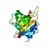
| ||||||||
|---|---|---|---|---|---|---|---|---|---|
| 1 |
| ||||||||
| Unit cell |
|
-Components
| #1: Protein | Mass: 14940.021 Da / Num. of mol.: 1 Source method: isolated from a genetically manipulated source Details: CRYSTALLIZED FROM AMMONIUM SULFATE SOLUTION / Source: (gene. exp.) Homo sapiens (human) / Tissue: PLATELET / Production host:  |
|---|---|
| #2: Chemical | ChemComp-SO4 /   Mass: 96.063 Da / Num. of mol.: 1 / Source method: obtained synthetically / Formula: SO4 Mass: 96.063 Da / Num. of mol.: 1 / Source method: obtained synthetically / Formula: SO4 |
| #3: Water | ChemComp-HOH /  Mass: 18.015 Da / Num. of mol.: 47 / Source method: isolated from a natural source / Formula: H2O Mass: 18.015 Da / Num. of mol.: 47 / Source method: isolated from a natural source / Formula: H2O |
-Experimental details
-Experiment
| Experiment | Method: X-RAY DIFFRACTION / Number of used crystals: 1 |
|---|
- Sample preparation
| Crystal | Density Matthews: 2.09 Å3/Da / Density % sol: 59 % |
|---|---|
| Crystal grow | pH: 6.7 Details: CRYSTALLIZED FROM AMMONIUM SULFATE SOLUTION, pH 6.7 |
-Data collection
| Diffraction | Mean temperature: 290 K |
|---|---|
| Diffraction source | Source: ROTATING ANODE / Type: RIGAKU RUH2R / Wavelength: 1.5418 |
| Detector | Type: SIEMENS-NICOLET X100 / Detector: AREA DETECTOR / Date: 1993 |
| Radiation | Monochromator: GRAPHITE(002) / Monochromatic (M) / Laue (L): M / Scattering type: x-ray |
| Radiation wavelength | Wavelength: 1.5418 Å / Relative weight: 1 |
| Reflection | Resolution: 2.3→28.3 Å / Num. obs: 8076 / % possible obs: 94.1 % / Observed criterion σ(I): 2 / Redundancy: 1.28 % / Rmerge(I) obs: 0.041 / Net I/σ(I): 18.2 |
- Processing
| Software |
| ||||||||||||||||||||||||||||||||||||||||||||||||||||||||||||||||||||||||||||||||||||
|---|---|---|---|---|---|---|---|---|---|---|---|---|---|---|---|---|---|---|---|---|---|---|---|---|---|---|---|---|---|---|---|---|---|---|---|---|---|---|---|---|---|---|---|---|---|---|---|---|---|---|---|---|---|---|---|---|---|---|---|---|---|---|---|---|---|---|---|---|---|---|---|---|---|---|---|---|---|---|---|---|---|---|---|---|---|
| Refinement | Method to determine structure: molecular replacement Starting model: 1FIL Resolution: 2→8 Å / σ(F): 2
| ||||||||||||||||||||||||||||||||||||||||||||||||||||||||||||||||||||||||||||||||||||
| Refine analyze | Luzzati coordinate error obs: 0.2 Å | ||||||||||||||||||||||||||||||||||||||||||||||||||||||||||||||||||||||||||||||||||||
| Refinement step | Cycle: LAST / Resolution: 2→8 Å
| ||||||||||||||||||||||||||||||||||||||||||||||||||||||||||||||||||||||||||||||||||||
| Refine LS restraints |
|
