 Movie
Movie Controller

[English] 日本語
 Yorodumi
- PDB-3p95: Human mesotrypsin complexed with bovine pancreatic trypsin inhibi... -
+ Open data

- Basic information
| Entry | Database: PDB / ID: 3p95 | ||||||
|---|---|---|---|---|---|---|---|
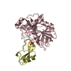
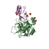
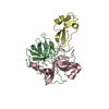
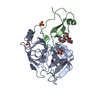
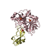
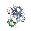
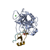
| Title | Human mesotrypsin complexed with bovine pancreatic trypsin inhibitor variant (BPTI-K15R/R17D) | ||||||
 Components Components |
| ||||||
 Keywords Keywords | HYDROLASE/HYDROLASE INHIBITOR / Human mesotrypsin-canonical inhibitor complex / Mesotrypsin / Trypsin IV / Bovine Pancreatic Trypsin Inhibitor / canonical inhibitor / BPTI-K15R/R17D / HYDROLASE-HYDROLASE INHIBITOR complex | ||||||
| Function / homology |  Function and homology information Function and homology informationUptake of dietary cobalamins into enterocytes / sulfate binding / negative regulation of platelet aggregation / zymogen binding / antimicrobial humoral response / Alpha-defensins / potassium channel inhibitor activity / Differentiation of Keratinocytes in Interfollicular Epidermis in Mammalian Skin / Antimicrobial peptides / zymogen activation ...Uptake of dietary cobalamins into enterocytes / sulfate binding / negative regulation of platelet aggregation / zymogen binding / antimicrobial humoral response / Alpha-defensins / potassium channel inhibitor activity / Differentiation of Keratinocytes in Interfollicular Epidermis in Mammalian Skin / Antimicrobial peptides / zymogen activation / molecular function inhibitor activity / negative regulation of thrombin-activated receptor signaling pathway / trypsin / endothelial cell migration / serine protease inhibitor complex / digestion / serine-type peptidase activity / serine-type endopeptidase inhibitor activity / tertiary granule lumen / protease binding / serine-type endopeptidase activity / calcium ion binding / Neutrophil degranulation / proteolysis / : / extracellular region / metal ion binding Similarity search - Function | ||||||
| Biological species |  Homo sapiens (human) Homo sapiens (human) | ||||||
| Method |  X-RAY DIFFRACTION / SYNCHROTRON / MOLECULAR REPLACEMENT / molecular replacement / Resolution: 1.2991 Å X-RAY DIFFRACTION / SYNCHROTRON / MOLECULAR REPLACEMENT / molecular replacement / Resolution: 1.2991 Å | ||||||
 Authors Authors | Salameh, M.A. / Soares, A.S. / Radisky, E.S. | ||||||
 Citation Citation | Journal: Biochem.J. / Year: 2011 Title: The P2' residue is a key determinant of mesotrypsin specificity: engineering a high-affinity inhibitor with anticancer activity. Authors: Salameh, M.A. / Soares, A.S. / Hockla, A. / Radisky, D.C. / Radisky, E.S. | ||||||
| History |
|
- Structure visualization
| Structure viewer | Molecule: MolmilJmol/JSmol |
|---|
- Downloads & links
-Download
| PDBx/mmCIF format | 3p95.cif.gz | 181.6 KB | Display | PDBx/mmCIF format |
|---|---|---|---|---|
| PDB format | pdb3p95.ent.gz | 145.3 KB | Display | PDB format |
| PDBx/mmJSON format | 3p95.json.gz | Tree view | PDBx/mmJSON format | |
| Others |  Other downloads Other downloads |
-Validation report
| Arichive directory | https://data.pdbj.org/pub/pdb/validation_reports/p9/3p95ftp://data.pdbj.org/pub/pdb/validation_reports/p9/3p95 | HTTPS FTP |
|---|
-Related structure data
| Related structure data |  3p92C  2r9pS S: Starting model for refinement C: citing same article ( |
|---|---|
| Similar structure data |


-Links
 PDBj
PDBj


- Assembly
| Deposited unit | 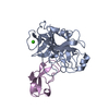
| ||||||||
|---|---|---|---|---|---|---|---|---|---|
| 1 |
| ||||||||
| Unit cell |
|
-Components
| #1: Protein | Mass: 24257.457 Da / Num. of mol.: 1 / Mutation: S195A Source method: isolated from a genetically manipulated source Source: (gene. exp.) Homo sapiens (human) / Gene: PRSS3 / Production host:  |
|---|---|
| #2: Protein | Mass: 6513.476 Da / Num. of mol.: 1 / Fragment: UNP Residues 36-93 / Mutation: K15R, R17D Source method: isolated from a genetically manipulated source Source: (gene. exp.)  PICHIA PASTORIS (fungus) / References: UniProt: P00974 PICHIA PASTORIS (fungus) / References: UniProt: P00974 |
| #3: Chemical | ChemComp-CA /   Mass: 40.078 Da / Num. of mol.: 1 / Source method: obtained synthetically / Formula: Ca Mass: 40.078 Da / Num. of mol.: 1 / Source method: obtained synthetically / Formula: Ca |
| #4: Water | ChemComp-HOH /  Mass: 18.015 Da / Num. of mol.: 280 / Source method: isolated from a natural source / Formula: H2O Mass: 18.015 Da / Num. of mol.: 280 / Source method: isolated from a natural source / Formula: H2O |
| Has protein modification | Y |
-Experimental details
-Experiment
| Experiment | Method: X-RAY DIFFRACTION / Number of used crystals: 1 |
|---|
- Sample preparation
| Crystal | Density Matthews: 1.88 Å3/Da / Density % sol: 34.58 % |
|---|---|
| Crystal grow | Temperature: 298 K / Method: vapor diffusion, hanging drop / pH: 8 Details: 25% PEG4000, 0.2M Na acetate and 100mM Tris pH 8.0, vapor diffusion, hanging drop, temperature 298K |
-Data collection
| Diffraction source | Source: SYNCHROTRON / Site: NSLS  / Beamline: X12B / Beamline: X12B | |||||||||||||||||||||||||||||||||||||||||||||||||||||||||||||||||||||||||||||||||||||||||||||||||||||||||||||||||||||||||||||||||||||||||||||||||||
|---|---|---|---|---|---|---|---|---|---|---|---|---|---|---|---|---|---|---|---|---|---|---|---|---|---|---|---|---|---|---|---|---|---|---|---|---|---|---|---|---|---|---|---|---|---|---|---|---|---|---|---|---|---|---|---|---|---|---|---|---|---|---|---|---|---|---|---|---|---|---|---|---|---|---|---|---|---|---|---|---|---|---|---|---|---|---|---|---|---|---|---|---|---|---|---|---|---|---|---|---|---|---|---|---|---|---|---|---|---|---|---|---|---|---|---|---|---|---|---|---|---|---|---|---|---|---|---|---|---|---|---|---|---|---|---|---|---|---|---|---|---|---|---|---|---|---|---|---|
| Detector | Type: ADSC QUANTUM 4 / Detector: CCD | |||||||||||||||||||||||||||||||||||||||||||||||||||||||||||||||||||||||||||||||||||||||||||||||||||||||||||||||||||||||||||||||||||||||||||||||||||
| Radiation | Protocol: SINGLE WAVELENGTH / Monochromatic (M) / Laue (L): M / Scattering type: x-ray | |||||||||||||||||||||||||||||||||||||||||||||||||||||||||||||||||||||||||||||||||||||||||||||||||||||||||||||||||||||||||||||||||||||||||||||||||||
| Radiation wavelength | Relative weight: 1 | |||||||||||||||||||||||||||||||||||||||||||||||||||||||||||||||||||||||||||||||||||||||||||||||||||||||||||||||||||||||||||||||||||||||||||||||||||
| Reflection | Resolution: 1.299→50 Å / Num. obs: 53441 / % possible obs: 94.4 % / Redundancy: 6.3 % / Rmerge(I) obs: 0.068 / Χ2: 1.21 / Net I/σ(I): 17.1 | |||||||||||||||||||||||||||||||||||||||||||||||||||||||||||||||||||||||||||||||||||||||||||||||||||||||||||||||||||||||||||||||||||||||||||||||||||
| Reflection shell | Diffraction-ID: 1
|
-Phasing
| Phasing | Method: molecular replacement |
|---|
- Processing
| Software |
| |||||||||||||||||||||||||||||||||||||||||||||||||||||||||||||||||||||||||||||||||||||||||||||||||||||||||
|---|---|---|---|---|---|---|---|---|---|---|---|---|---|---|---|---|---|---|---|---|---|---|---|---|---|---|---|---|---|---|---|---|---|---|---|---|---|---|---|---|---|---|---|---|---|---|---|---|---|---|---|---|---|---|---|---|---|---|---|---|---|---|---|---|---|---|---|---|---|---|---|---|---|---|---|---|---|---|---|---|---|---|---|---|---|---|---|---|---|---|---|---|---|---|---|---|---|---|---|---|---|---|---|---|---|---|
| Refinement | Method to determine structure: MOLECULAR REPLACEMENT Starting model: PDB ENTRY 2R9P Resolution: 1.2991→33.817 Å / Occupancy max: 1 / Occupancy min: 0 / FOM work R set: 0.9487 / SU ML: 0.07 / σ(F): 0.06 / Phase error: 10.34 / Stereochemistry target values: MLHL
| |||||||||||||||||||||||||||||||||||||||||||||||||||||||||||||||||||||||||||||||||||||||||||||||||||||||||
| Solvent computation | Shrinkage radii: 0.9 Å / VDW probe radii: 1.11 Å / Solvent model: FLAT BULK SOLVENT MODEL / Bsol: 62.547 Å2 / ksol: 0.485 e/Å3 | |||||||||||||||||||||||||||||||||||||||||||||||||||||||||||||||||||||||||||||||||||||||||||||||||||||||||
| Displacement parameters | Biso max: 119.66 Å2 / Biso mean: 19.2292 Å2 / Biso min: 6.32 Å2
| |||||||||||||||||||||||||||||||||||||||||||||||||||||||||||||||||||||||||||||||||||||||||||||||||||||||||
| Refinement step | Cycle: LAST / Resolution: 1.2991→33.817 Å
| |||||||||||||||||||||||||||||||||||||||||||||||||||||||||||||||||||||||||||||||||||||||||||||||||||||||||
| Refine LS restraints |
| |||||||||||||||||||||||||||||||||||||||||||||||||||||||||||||||||||||||||||||||||||||||||||||||||||||||||
| LS refinement shell | Refine-ID: X-RAY DIFFRACTION / Total num. of bins used: 14
|
