 Movie
Movie Controller

[English] 日本語
 Yorodumi
- PDB-3p39: Crystal structure of the NS1 effector domain W182A mutant from in... -
+ Open data

- Basic information
| Entry | Database: PDB / ID: 3p39 | ||||||
|---|---|---|---|---|---|---|---|
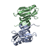
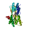
| Title | Crystal structure of the NS1 effector domain W182A mutant from influenza A/Vietnam/1203/2004 (H5N1) virus | ||||||
 Components Components | Nonstructural protein 1 | ||||||
 Keywords Keywords | VIRAL PROTEIN | ||||||
| Function / homology |  Function and homology information Function and homology informationsymbiont-mediated suppression of host mRNA processing / symbiont-mediated suppression of host PKR/eIFalpha signaling / symbiont-mediated suppression of host cytoplasmic pattern recognition receptor signaling pathway via inhibition of RIG-I activity / protein serine/threonine kinase inhibitor activity / host cell cytoplasm / symbiont-mediated suppression of host type I interferon-mediated signaling pathway / symbiont-mediated suppression of host gene expression / host cell nucleus / RNA binding Similarity search - Function | ||||||
| Biological species |   Influenza A virus Influenza A virus | ||||||
| Method |  X-RAY DIFFRACTION / SYNCHROTRON / MOLECULAR REPLACEMENT / Resolution: 3.14 Å X-RAY DIFFRACTION / SYNCHROTRON / MOLECULAR REPLACEMENT / Resolution: 3.14 Å | ||||||
 Authors Authors | Chen, S. / Xiao, Y.B. / Bricogne, G. / Sharff, A.J. / Hilgenfeld, R. | ||||||
 Citation Citation | Journal: To be Published Title: X-ray structures of the NS1 effector domain from highly pathogenic influenza A/Vietnam/1203/2004 (H5N1) virus Authors: Chen, S. / Xiao, Y.B. / Bricogne, G. / Sharff, A.J. / Hilgenfeld, R. | ||||||
| History |
|
- Structure visualization
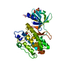
| Structure viewer | Molecule: MolmilJmol/JSmol |
|---|
- Downloads & links
-Download
| PDBx/mmCIF format | 3p39.cif.gz | 154.8 KB | Display | PDBx/mmCIF format |
|---|---|---|---|---|
| PDB format | pdb3p39.ent.gz | 122.8 KB | Display | PDB format |
| PDBx/mmJSON format | 3p39.json.gz | Tree view | PDBx/mmJSON format | |
| Others |  Other downloads Other downloads |
-Validation report
| Arichive directory | https://data.pdbj.org/pub/pdb/validation_reports/p3/3p39ftp://data.pdbj.org/pub/pdb/validation_reports/p3/3p39 | HTTPS FTP |
|---|
-Related structure data
| Related structure data |  3p31SC  3p38C S: Starting model for refinement C: citing same article ( |
|---|---|
| Similar structure data |
-Links
 PDBj
PDBj- Assembly
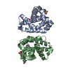
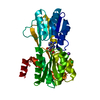
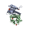
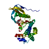
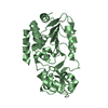
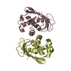
| Deposited unit | 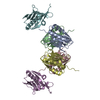
| ||||||||
|---|---|---|---|---|---|---|---|---|---|
| 1 | 
| ||||||||
| 2 | 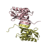
| ||||||||
| 3 | 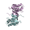
| ||||||||
| 4 | 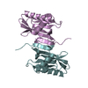
| ||||||||
| Unit cell |
|
-Components
| #1: Protein | Mass: 16346.924 Da / Num. of mol.: 6 / Fragment: C-terminal effector domain, UNP residues 73-215 / Mutation: W182A Source method: isolated from a genetically manipulated source Source: (gene. exp.) Influenza A virus / Strain: A/environment/Viet Nam/1203/2004(H5N1) / Gene: NS1 / Plasmid: pGEX6P-1 / Production host:  #2: Water | ChemComp-HOH / |  Mass: 18.015 Da / Num. of mol.: 100 / Source method: isolated from a natural source / Formula: H2O Mass: 18.015 Da / Num. of mol.: 100 / Source method: isolated from a natural source / Formula: H2O |
|---|
-Experimental details
-Experiment
| Experiment | Method: X-RAY DIFFRACTION / Number of used crystals: 1 |
|---|
- Sample preparation
| Crystal | Density Matthews: 3.24 Å3/Da / Density % sol: 62.05 % |
|---|---|
| Crystal grow | Temperature: 285 K / Method: vapor diffusion, sitting drop / pH: 8.4 Details: 0.1M Bicine, 1.8 M MgCl2, pH 8.4, VAPOR DIFFUSION, SITTING DROP, temperature 285K |
-Data collection
| Diffraction | Mean temperature: 100 K |
|---|---|
| Diffraction source | Source: SYNCHROTRON / Site: BESSY  / Beamline: 14.1 / Wavelength: 0.9184 Å / Beamline: 14.1 / Wavelength: 0.9184 Å |
| Detector | Type: RAYONIX MX-225 / Detector: CCD / Date: Feb 19, 2010 |
| Radiation | Monochromator: Si 111 Crystal / Protocol: SINGLE WAVELENGTH / Monochromatic (M) / Laue (L): M / Scattering type: x-ray |
| Radiation wavelength | Wavelength: 0.9184 Å / Relative weight: 1 |
| Reflection | Resolution: 3.14→37.3 Å / Num. all: 22045 / Num. obs: 21988 / % possible obs: 94.61 % / Observed criterion σ(F): 0 / Observed criterion σ(I): 0 / Redundancy: 7.3 % / Biso Wilson estimate: 51.83 Å2 / Rmerge(I) obs: 0.109 / Net I/σ(I): 16.8 |
| Reflection shell | Resolution: 3.14→3.29 Å / Redundancy: 1.7 % / Rmerge(I) obs: 0.272 / Mean I/σ(I) obs: 2.2 / % possible all: 46.5 |
- Processing
| Software |
| ||||||||||||||||||||||||||||||||||||||||||||||||||||||||||||||||||||||||||||||||||||||||||||||||||||||||||||
|---|---|---|---|---|---|---|---|---|---|---|---|---|---|---|---|---|---|---|---|---|---|---|---|---|---|---|---|---|---|---|---|---|---|---|---|---|---|---|---|---|---|---|---|---|---|---|---|---|---|---|---|---|---|---|---|---|---|---|---|---|---|---|---|---|---|---|---|---|---|---|---|---|---|---|---|---|---|---|---|---|---|---|---|---|---|---|---|---|---|---|---|---|---|---|---|---|---|---|---|---|---|---|---|---|---|---|---|---|---|
| Refinement | Method to determine structure: MOLECULAR REPLACEMENT Starting model: PDB ENTRY 3P31 Resolution: 3.14→37.3 Å / Cor.coef. Fo:Fc: 0.8882 / Cor.coef. Fo:Fc free: 0.856 / Occupancy max: 1 / Occupancy min: 1 / Cross valid method: THROUGHOUT / σ(F): 0 / Stereochemistry target values: Engh & Huber
| ||||||||||||||||||||||||||||||||||||||||||||||||||||||||||||||||||||||||||||||||||||||||||||||||||||||||||||
| Displacement parameters | Biso mean: 47.86 Å2
| ||||||||||||||||||||||||||||||||||||||||||||||||||||||||||||||||||||||||||||||||||||||||||||||||||||||||||||
| Refine analyze | Luzzati coordinate error obs: 0.614 Å | ||||||||||||||||||||||||||||||||||||||||||||||||||||||||||||||||||||||||||||||||||||||||||||||||||||||||||||
| Refinement step | Cycle: LAST / Resolution: 3.14→37.3 Å
| ||||||||||||||||||||||||||||||||||||||||||||||||||||||||||||||||||||||||||||||||||||||||||||||||||||||||||||
| Refine LS restraints |
| ||||||||||||||||||||||||||||||||||||||||||||||||||||||||||||||||||||||||||||||||||||||||||||||||||||||||||||
| LS refinement shell | Resolution: 3.14→3.29 Å / Total num. of bins used: 11
|
