 Movie
Movie Controller

[English] 日本語
 Yorodumi
- PDB-4ijx: Crystal structure of human Ap4A hydrolase E58A mutant complexed w... -
+ Open data

- Basic information
| Entry | Database: PDB / ID: 4ijx | ||||||
|---|---|---|---|---|---|---|---|
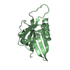
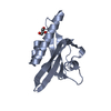
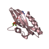
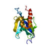
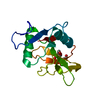
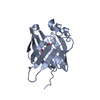
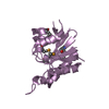
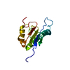
| Title | Crystal structure of human Ap4A hydrolase E58A mutant complexed with DPO | ||||||
 Components Components | Bis(5'-nucleosyl)-tetraphosphatase [asymmetrical] | ||||||
 Keywords Keywords | HYDROLASE / NUDIX FOLD | ||||||
| Function / homology |  Function and homology information Function and homology informationbis(5'-nucleosyl)-tetraphosphatase (asymmetrical) / AMP biosynthetic process / bis(5'-nucleosyl)-tetraphosphatase (symmetrical) activity / bis(5'-nucleosyl)-tetraphosphatase (asymmetrical) activity / ATP biosynthetic process / nucleobase-containing compound metabolic process / Detoxification of Reactive Oxygen Species / cellular response to oxidative stress / mitochondrial matrix / apoptotic process / GTP binding Similarity search - Function | ||||||
| Biological species |  Homo sapiens (human) Homo sapiens (human) | ||||||
| Method |  X-RAY DIFFRACTION / SYNCHROTRON / MOLECULAR REPLACEMENT / Resolution: 2.1 Å X-RAY DIFFRACTION / SYNCHROTRON / MOLECULAR REPLACEMENT / Resolution: 2.1 Å | ||||||
 Authors Authors | Ge, H. / Chen, X. | ||||||
 Citation Citation | Journal: Biochem.Biophys.Res.Commun. / Year: 2013 Title: Crystal structure of wild-type and mutant human Ap4A hydrolase. Authors: Ge, H. / Chen, X. / Yang, W. / Niu, L. / Teng, M. | ||||||
| History |
|
- Structure visualization
| Structure viewer | Molecule: MolmilJmol/JSmol |
|---|
- Downloads & links
-Download
| PDBx/mmCIF format | 4ijx.cif.gz | 134.9 KB | Display | PDBx/mmCIF format |
|---|---|---|---|---|
| PDB format | pdb4ijx.ent.gz | 106.5 KB | Display | PDB format |
| PDBx/mmJSON format | 4ijx.json.gz | Tree view | PDBx/mmJSON format | |
| Others |  Other downloads Other downloads |
-Validation report
| Arichive directory | https://data.pdbj.org/pub/pdb/validation_reports/ij/4ijxftp://data.pdbj.org/pub/pdb/validation_reports/ij/4ijx | HTTPS FTP |
|---|
-Related structure data
| Related structure data | 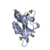 3u53SC  4ickC S: Starting model for refinement C: citing same article ( |
|---|---|
| Similar structure data |
-Links
 PDBj
PDBj


- Assembly
| Deposited unit | 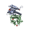
| ||||||||
|---|---|---|---|---|---|---|---|---|---|
| 1 | 
| ||||||||
| 2 | 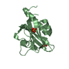
| ||||||||
| 3 | 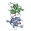
| ||||||||
| Unit cell |
|
-Components
-Protein , 1 types, 2 molecules AB
| #1: Protein | Mass: 17864.350 Da / Num. of mol.: 2 / Mutation: E58A Source method: isolated from a genetically manipulated source Source: (gene. exp.) Homo sapiens (human) / Gene: NUDT2, APAH1 / Plasmid: pET22b / Production host:  References: UniProt: P50583, bis(5'-nucleosyl)-tetraphosphatase (asymmetrical) |
|---|
-Non-polymers , 5 types, 126 molecules 


| #2: Chemical | ChemComp-MG /  Mass: 24.305 Da / Num. of mol.: 1 / Source method: obtained synthetically / Formula: Mg Mass: 24.305 Da / Num. of mol.: 1 / Source method: obtained synthetically / Formula: Mg | ||
|---|---|---|---|
| #3: Chemical | ChemComp-DPO /  Mass: 173.943 Da / Num. of mol.: 1 / Source method: obtained synthetically / Formula: O7P2 Mass: 173.943 Da / Num. of mol.: 1 / Source method: obtained synthetically / Formula: O7P2 | ||
| #4: Chemical | ChemComp-GOL /  Mass: 92.094 Da / Num. of mol.: 1 / Source method: obtained synthetically / Formula: C3H8O3 Mass: 92.094 Da / Num. of mol.: 1 / Source method: obtained synthetically / Formula: C3H8O3 | ||
| #5: Chemical |  Mass: 94.971 Da / Num. of mol.: 2 / Source method: obtained synthetically / Formula: PO4 Mass: 94.971 Da / Num. of mol.: 2 / Source method: obtained synthetically / Formula: PO4#6: Water | ChemComp-HOH / | Mass: 18.015 Da / Num. of mol.: 121 / Source method: isolated from a natural source / Formula: H2O |
-Experimental details
-Experiment
| Experiment | Method: X-RAY DIFFRACTION / Number of used crystals: 1 |
|---|
- Sample preparation
| Crystal | Density Matthews: 2.44 Å3/Da / Density % sol: 49.67 % |
|---|---|
| Crystal grow | Temperature: 285 K / Method: vapor diffusion, hanging drop / pH: 8.4 Details: 0.1M Tris, 2.0M Ammonium phosphate monobasic, 5mM magnesium chloride, pH 8.4, VAPOR DIFFUSION, HANGING DROP, temperature 285K |
-Data collection
| Diffraction | Mean temperature: 100 K |
|---|---|
| Diffraction source | Source: SYNCHROTRON / Site: SSRF  / Beamline: BL17U / Wavelength: 0.97915 Å / Beamline: BL17U / Wavelength: 0.97915 Å |
| Detector | Date: Jun 21, 2012 |
| Radiation | Protocol: SINGLE WAVELENGTH / Monochromatic (M) / Laue (L): M / Scattering type: x-ray |
| Radiation wavelength | Wavelength: 0.97915 Å / Relative weight: 1 |
| Reflection | Resolution: 2.1→30 Å / Num. all: 21348 / Num. obs: 20214 |
| Reflection shell | Resolution: 2.103→2.157 Å / Num. unique all: 1277 / % possible all: 98.9 |
- Processing
| Software |
| |||||||||||||||||||||||||||||||||||||||||||||||||||||||||||||||||||||||||||||||||||||||||||||||||||||||||||||||||||||||||||||||||||||||||||||||||||||||||||||||||||||||||||||||||||||||||||||||||||||||||||||||||||||||||||||||||
|---|---|---|---|---|---|---|---|---|---|---|---|---|---|---|---|---|---|---|---|---|---|---|---|---|---|---|---|---|---|---|---|---|---|---|---|---|---|---|---|---|---|---|---|---|---|---|---|---|---|---|---|---|---|---|---|---|---|---|---|---|---|---|---|---|---|---|---|---|---|---|---|---|---|---|---|---|---|---|---|---|---|---|---|---|---|---|---|---|---|---|---|---|---|---|---|---|---|---|---|---|---|---|---|---|---|---|---|---|---|---|---|---|---|---|---|---|---|---|---|---|---|---|---|---|---|---|---|---|---|---|---|---|---|---|---|---|---|---|---|---|---|---|---|---|---|---|---|---|---|---|---|---|---|---|---|---|---|---|---|---|---|---|---|---|---|---|---|---|---|---|---|---|---|---|---|---|---|---|---|---|---|---|---|---|---|---|---|---|---|---|---|---|---|---|---|---|---|---|---|---|---|---|---|---|---|---|---|---|---|---|---|---|---|---|---|---|---|---|---|---|---|---|---|---|---|---|
| Refinement | Method to determine structure: MOLECULAR REPLACEMENT Starting model: 3u53 Resolution: 2.1→30 Å / Cor.coef. Fo:Fc: 0.953 / Cor.coef. Fo:Fc free: 0.927 / SU B: 8.654 / SU ML: 0.115 / Cross valid method: THROUGHOUT / ESU R Free: 0.176 / Stereochemistry target values: MAXIMUM LIKELIHOOD / Details: HYDROGENS HAVE BEEN USED IF PRESENT IN THE INPUT
| |||||||||||||||||||||||||||||||||||||||||||||||||||||||||||||||||||||||||||||||||||||||||||||||||||||||||||||||||||||||||||||||||||||||||||||||||||||||||||||||||||||||||||||||||||||||||||||||||||||||||||||||||||||||||||||||||
| Solvent computation | Ion probe radii: 0.8 Å / Shrinkage radii: 0.8 Å / VDW probe radii: 1.2 Å / Solvent model: MASK | |||||||||||||||||||||||||||||||||||||||||||||||||||||||||||||||||||||||||||||||||||||||||||||||||||||||||||||||||||||||||||||||||||||||||||||||||||||||||||||||||||||||||||||||||||||||||||||||||||||||||||||||||||||||||||||||||
| Displacement parameters | Biso mean: 37.373 Å2
| |||||||||||||||||||||||||||||||||||||||||||||||||||||||||||||||||||||||||||||||||||||||||||||||||||||||||||||||||||||||||||||||||||||||||||||||||||||||||||||||||||||||||||||||||||||||||||||||||||||||||||||||||||||||||||||||||
| Refinement step | Cycle: LAST / Resolution: 2.1→30 Å
| |||||||||||||||||||||||||||||||||||||||||||||||||||||||||||||||||||||||||||||||||||||||||||||||||||||||||||||||||||||||||||||||||||||||||||||||||||||||||||||||||||||||||||||||||||||||||||||||||||||||||||||||||||||||||||||||||
| Refine LS restraints |
| |||||||||||||||||||||||||||||||||||||||||||||||||||||||||||||||||||||||||||||||||||||||||||||||||||||||||||||||||||||||||||||||||||||||||||||||||||||||||||||||||||||||||||||||||||||||||||||||||||||||||||||||||||||||||||||||||
| LS refinement shell | Resolution: 2.103→2.157 Å / Total num. of bins used: 20
| |||||||||||||||||||||||||||||||||||||||||||||||||||||||||||||||||||||||||||||||||||||||||||||||||||||||||||||||||||||||||||||||||||||||||||||||||||||||||||||||||||||||||||||||||||||||||||||||||||||||||||||||||||||||||||||||||
| Refinement TLS params. | Method: refined / Refine-ID: X-RAY DIFFRACTION
| |||||||||||||||||||||||||||||||||||||||||||||||||||||||||||||||||||||||||||||||||||||||||||||||||||||||||||||||||||||||||||||||||||||||||||||||||||||||||||||||||||||||||||||||||||||||||||||||||||||||||||||||||||||||||||||||||
| Refinement TLS group |
|
