 Movie
Movie Controller

[English] 日本語
 Yorodumi
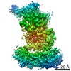
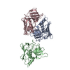
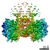
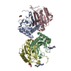
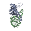
- PDB-3p30: crystal structure of the cluster II Fab 1281 in complex with HIV-... -
+ Open data

- Basic information
| Entry | Database: PDB / ID: 3p30 | ||||||
|---|---|---|---|---|---|---|---|
| Title | crystal structure of the cluster II Fab 1281 in complex with HIV-1 gp41 ectodomain | ||||||
 Components Components |
| ||||||
 Keywords Keywords | IMMUNE SYSTEM / HIV / gp41 / cluster 2 / Fab / 6-helix bundle | ||||||
| Function / homology | Helix Hairpins - #210 / Helix Hairpins / Immunoglobulins / Immunoglobulin-like / Sandwich / Orthogonal Bundle / Mainly Beta / Mainly Alpha Function and homology information Function and homology information | ||||||
| Biological species |   Human immunodeficiency virus 1 Human immunodeficiency virus 1 Homo sapiens (human) Homo sapiens (human) | ||||||
| Method |  X-RAY DIFFRACTION / SYNCHROTRON / MOLECULAR REPLACEMENT / Resolution: 3.3 Å X-RAY DIFFRACTION / SYNCHROTRON / MOLECULAR REPLACEMENT / Resolution: 3.3 Å | ||||||
 Authors Authors | Frey, G. / Chen, J. / Rits-Volloch, S. / Freeman, M.M. / Zolla-Pazner, S. / Chen, B. | ||||||
 Citation Citation | Journal: Nat.Struct.Mol.Biol. / Year: 2010 Title: Distinct conformational states of HIV-1 gp41 are recognized by neutralizing and non-neutralizing antibodies. Authors: Frey, G. / Chen, J. / Rits-Volloch, S. / Freeman, M.M. / Zolla-Pazner, S. / Chen, B. | ||||||
| History |
|
- Structure visualization
| Structure viewer | Molecule: MolmilJmol/JSmol |
|---|
- Downloads & links
-Download
| PDBx/mmCIF format | 3p30.cif.gz | 210.1 KB | Display | PDBx/mmCIF format |
|---|---|---|---|---|
| PDB format | pdb3p30.ent.gz | 169.4 KB | Display | PDB format |
| PDBx/mmJSON format | 3p30.json.gz | Tree view | PDBx/mmJSON format | |
| Others |  Other downloads Other downloads |
-Validation report
| Summary document | 3p30_validation.pdf.gz | 449.1 KB | Display | wwPDB validaton report |
|---|---|---|---|---|
| Full document | 3p30_full_validation.pdf.gz | 452.7 KB | Display | |
| Data in XML | 3p30_validation.xml.gz | 18.9 KB | Display | |
| Data in CIF | 3p30_validation.cif.gz | 25.2 KB | Display | |
| Arichive directory | https://data.pdbj.org/pub/pdb/validation_reports/p3/3p30ftp://data.pdbj.org/pub/pdb/validation_reports/p3/3p30 | HTTPS FTP |
-Related structure data
| Similar structure data |
|---|


-Links
 PDBj
PDBj


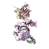
- Assembly
| Deposited unit | 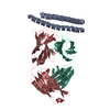
| ||||||||
|---|---|---|---|---|---|---|---|---|---|
| 1 |
| ||||||||
| Unit cell |
|
-Components
| #1: Protein | Mass: 11177.756 Da / Num. of mol.: 1 Source method: isolated from a genetically manipulated source Source: (gene. exp.) Human immunodeficiency virus 1 / Gene: HIV-1 ENV / Plasmid: pET21a / Production host:  |
|---|---|
| #2: Antibody | Mass: 22616.062 Da / Num. of mol.: 1 Source method: isolated from a genetically manipulated source Source: (gene. exp.) Homo sapiens (human) / Cell line (production host): HYBRIDOMA CELL / Production host: homo sapiens (human) |
| #3: Antibody | Mass: 23983.721 Da / Num. of mol.: 1 Source method: isolated from a genetically manipulated source Source: (gene. exp.) Homo sapiens (human) / Cell line (production host): HYBRIDOMA CELL / Production host: homo sapiens (human) |
| Has protein modification | Y |
| Sequence details | AUTHORS STATE THAT THE GEN BANK ACCESSION CODE FOR 'HIV-1 ENV' 92UG037.8 (HIV1, SAMPLE 037 CLONE 08 ...AUTHORS STATE THAT THE GEN BANK ACCESSION CODE FOR 'HIV-1 ENV' 92UG037.8 (HIV1, SAMPLE 037 CLONE 08 FROM UGANDA (HIV192UG037WHO.01083hED) IS U09127 |
-Experimental details
-Experiment
| Experiment | Method: X-RAY DIFFRACTION / Number of used crystals: 1 |
|---|
- Sample preparation
| Crystal | Density Matthews: 2.73 Å3/Da / Density % sol: 55.01 % |
|---|---|
| Crystal grow | Temperature: 298 K / Method: vapor diffusion, hanging drop / pH: 8 Details: PEG 4000, pH 8.0, VAPOR DIFFUSION, HANGING DROP, temperature 298K |
-Data collection
| Diffraction | Mean temperature: 100 K |
|---|---|
| Diffraction source | Source: SYNCHROTRON / Site: APS  / Beamline: 24-ID-E / Wavelength: 0.97916 / Beamline: 24-ID-E / Wavelength: 0.97916 |
| Detector | Type: ADSC QUANTUM 315 / Detector: CCD / Date: Mar 18, 2010 |
| Radiation | Monochromator: CRYOGENICALLY-COOLED SINGLE CRYSTAL SI(111) SIDE BOUNCE MONOCHROMATOR Protocol: SINGLE WAVELENGTH / Scattering type: x-ray |
| Radiation wavelength | Wavelength: 0.97916 Å / Relative weight: 1 |
| Reflection | Resolution: 3.3→33.44 Å / Num. obs: 8478 / % possible obs: 99.5 % / Observed criterion σ(I): 2 / Redundancy: 3.2 % / Rmerge(I) obs: 0.087 / Net I/σ(I): 9.5 |
| Reflection shell | Resolution: 3.31→3.45 Å / Redundancy: 3.2 % / Rmerge(I) obs: 0.425 / % possible all: 99.9 |
- Processing
| Software |
| ||||||||||||||||||||||||||||||||||||||||||||||||||||||||||||||||||||||||||||||||||||||||||||||||||||||||||||||||||||||||||||||||||||||||||||||||||||||||||||||||||||||||||
|---|---|---|---|---|---|---|---|---|---|---|---|---|---|---|---|---|---|---|---|---|---|---|---|---|---|---|---|---|---|---|---|---|---|---|---|---|---|---|---|---|---|---|---|---|---|---|---|---|---|---|---|---|---|---|---|---|---|---|---|---|---|---|---|---|---|---|---|---|---|---|---|---|---|---|---|---|---|---|---|---|---|---|---|---|---|---|---|---|---|---|---|---|---|---|---|---|---|---|---|---|---|---|---|---|---|---|---|---|---|---|---|---|---|---|---|---|---|---|---|---|---|---|---|---|---|---|---|---|---|---|---|---|---|---|---|---|---|---|---|---|---|---|---|---|---|---|---|---|---|---|---|---|---|---|---|---|---|---|---|---|---|---|---|---|---|---|---|---|---|---|---|
| Refinement | Method to determine structure: MOLECULAR REPLACEMENT / Resolution: 3.3→33.44 Å / Cor.coef. Fo:Fc: 0.902 / Cor.coef. Fo:Fc free: 0.89 / Occupancy max: 1 / Occupancy min: 1 / SU B: 93.97 / SU ML: 0.654 / Cross valid method: THROUGHOUT / σ(F): 0 / ESU R Free: 0.65 / Stereochemistry target values: MAXIMUM LIKELIHOOD / Details: HYDROGENS HAVE BEEN ADDED IN THE RIDING
| ||||||||||||||||||||||||||||||||||||||||||||||||||||||||||||||||||||||||||||||||||||||||||||||||||||||||||||||||||||||||||||||||||||||||||||||||||||||||||||||||||||||||||
| Solvent computation | Ion probe radii: 0.8 Å / Shrinkage radii: 0.8 Å / VDW probe radii: 1.4 Å / Solvent model: MASK | ||||||||||||||||||||||||||||||||||||||||||||||||||||||||||||||||||||||||||||||||||||||||||||||||||||||||||||||||||||||||||||||||||||||||||||||||||||||||||||||||||||||||||
| Displacement parameters | Biso mean: 130.63 Å2
| ||||||||||||||||||||||||||||||||||||||||||||||||||||||||||||||||||||||||||||||||||||||||||||||||||||||||||||||||||||||||||||||||||||||||||||||||||||||||||||||||||||||||||
| Refinement step | Cycle: LAST / Resolution: 3.3→33.44 Å
| ||||||||||||||||||||||||||||||||||||||||||||||||||||||||||||||||||||||||||||||||||||||||||||||||||||||||||||||||||||||||||||||||||||||||||||||||||||||||||||||||||||||||||
| Refine LS restraints |
| ||||||||||||||||||||||||||||||||||||||||||||||||||||||||||||||||||||||||||||||||||||||||||||||||||||||||||||||||||||||||||||||||||||||||||||||||||||||||||||||||||||||||||
| LS refinement shell | Resolution: 3.3→3.38 Å / Total num. of bins used: 20
| ||||||||||||||||||||||||||||||||||||||||||||||||||||||||||||||||||||||||||||||||||||||||||||||||||||||||||||||||||||||||||||||||||||||||||||||||||||||||||||||||||||||||||
| Refinement TLS params. | Method: refined / Refine-ID: X-RAY DIFFRACTION
| ||||||||||||||||||||||||||||||||||||||||||||||||||||||||||||||||||||||||||||||||||||||||||||||||||||||||||||||||||||||||||||||||||||||||||||||||||||||||||||||||||||||||||
| Refinement TLS group |
|
