 Movie
Movie Controller

[English] 日本語
 Yorodumi
- PDB-3oyv: Crystal structure of an imelysin peptidase (BACOVA_03801) from Ba... -
+ Open data

- Basic information
| Entry | Database: PDB / ID: 3oyv | ||||||
|---|---|---|---|---|---|---|---|
| Title | Crystal structure of an imelysin peptidase (BACOVA_03801) from Bacteroides ovatus ATCC 8483 at 1.25 A resolution | ||||||
 Components Components | Imelysin | ||||||
 Keywords Keywords | HYDROLASE / OUTER MEMBRANE PROTEIN / EXTRACELLULAR ACTIVE SITE / METAL BINDING PROTEIN / STRUCTURAL GENOMICS / JOINT CENTER FOR STRUCTURAL GENOMICS / JCSG / PROTEIN STRUCTURE INITIATIVE / PSI-BIOLOGY | ||||||
| Function / homology | Imelysin-like domain / Imelysin-like domain superfamily / Imelysin / Prokaryotic membrane lipoprotein lipid attachment site profile. / Unknown ligand / Imelysin Function and homology information Function and homology information | ||||||
| Biological species |  Bacteroides ovatus ATCC 8483 (bacteria) Bacteroides ovatus ATCC 8483 (bacteria) | ||||||
| Method |  X-RAY DIFFRACTION / SYNCHROTRON / MAD / Resolution: 1.25 Å X-RAY DIFFRACTION / SYNCHROTRON / MAD / Resolution: 1.25 Å | ||||||
 Authors Authors | Joint Center for Structural Genomics (JCSG) | ||||||
 Citation Citation | Journal: Plos One / Year: 2011 Title: Structural and sequence analysis of imelysin-like proteins implicated in bacterial iron uptake. Authors: Xu, Q. / Rawlings, N.D. / Farr, C.L. / Chiu, H.J. / Grant, J.C. / Jaroszewski, L. / Klock, H.E. / Knuth, M.W. / Miller, M.D. / Weekes, D. / Elsliger, M.A. / Deacon, A.M. / Godzik, A. / ...Authors: Xu, Q. / Rawlings, N.D. / Farr, C.L. / Chiu, H.J. / Grant, J.C. / Jaroszewski, L. / Klock, H.E. / Knuth, M.W. / Miller, M.D. / Weekes, D. / Elsliger, M.A. / Deacon, A.M. / Godzik, A. / Lesley, S.A. / Wilson, I.A. | ||||||
| History |
|
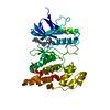
- Structure visualization
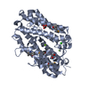
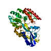
| Structure viewer | Molecule: MolmilJmol/JSmol |
|---|
- Downloads & links
-Download
| PDBx/mmCIF format | 3oyv.cif.gz | 189.9 KB | Display | PDBx/mmCIF format |
|---|---|---|---|---|
| PDB format | pdb3oyv.ent.gz | 150.5 KB | Display | PDB format |
| PDBx/mmJSON format | 3oyv.json.gz | Tree view | PDBx/mmJSON format | |
| Others |  Other downloads Other downloads |
-Validation report
| Arichive directory | https://data.pdbj.org/pub/pdb/validation_reports/oy/3oyvftp://data.pdbj.org/pub/pdb/validation_reports/oy/3oyv | HTTPS FTP |
|---|
-Related structure data


-Links
 PDBj
PDBj


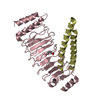
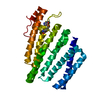
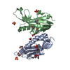
- Assembly
| Deposited unit | 
| ||||||||
|---|---|---|---|---|---|---|---|---|---|
| 1 |
| ||||||||
| Unit cell |
| ||||||||
| Components on special symmetry positions |
| ||||||||
| Details | CRYSTAL PACKING SUGGESTS THE ASSIGNMENT OF A MONOMER AS THE SIGNIFICANT OLIGOMERIZATION STATE IN SOLUTION. ANALYTICAL SIZE EXCLUSION CHROMATOGRAPHY WITH STATIC LIGHT SCATTERING PROVIDES SUPPORTING EVIDENCE THAT A MONOMER IS A SIGNIFICANT OLIGOMERIZATION STATE. |
-Components
| #1: Protein | Mass: 39963.996 Da / Num. of mol.: 1 / Fragment: sequence database residues 25-384 Source method: isolated from a genetically manipulated source Source: (gene. exp.) Bacteroides ovatus ATCC 8483 (bacteria)Gene: BACOVA_03801 / Plasmid: SpeedET / Production host: | ||||||
|---|---|---|---|---|---|---|---|
| #2: Chemical | ChemComp-UNL / Num. of mol.: 1 / Source method: obtained synthetically | ||||||
| #3: Chemical | ChemComp-CL /   Mass: 35.453 Da / Num. of mol.: 1 / Source method: obtained synthetically / Formula: Cl Mass: 35.453 Da / Num. of mol.: 1 / Source method: obtained synthetically / Formula: Cl | ||||||
| #4: Chemical |   Mass: 92.094 Da / Num. of mol.: 2 / Source method: obtained synthetically / Formula: C3H8O3 Mass: 92.094 Da / Num. of mol.: 2 / Source method: obtained synthetically / Formula: C3H8O3#5: Water | ChemComp-HOH / |  Mass: 18.015 Da / Num. of mol.: 533 / Source method: isolated from a natural source / Formula: H2O Mass: 18.015 Da / Num. of mol.: 533 / Source method: isolated from a natural source / Formula: H2OHas protein modification | Y | Sequence details | THE CONSTRUCT (RESIDUES 25-384) WAS EXPRESSED WITH A PURIFICATION TAG MGSDKIHHHHHHENLYFQG. THE TAG ...THE CONSTRUCT (RESIDUES 25-384) WAS EXPRESSED WITH A PURIFICATI | |
-Experimental details
-Experiment
| Experiment | Method: X-RAY DIFFRACTION / Number of used crystals: 1 |
|---|
- Sample preparation
| Crystal | Density Matthews: 2.16 Å3/Da / Density % sol: 43.1 % |
|---|---|
| Crystal grow | Temperature: 277 K / Method: vapor diffusion, sitting drop / pH: 4.5 Details: 0.05M KH2PO4, 20.00% PEG-8000, No Buffer pH 4.5, NANODROP, VAPOR DIFFUSION, SITTING DROP, temperature 277K |
-Data collection
| Diffraction | Mean temperature: 100 K | |||||||||||||||||||||||||||||||||||||||||||||||||||||||||||||||||||||||||||||
|---|---|---|---|---|---|---|---|---|---|---|---|---|---|---|---|---|---|---|---|---|---|---|---|---|---|---|---|---|---|---|---|---|---|---|---|---|---|---|---|---|---|---|---|---|---|---|---|---|---|---|---|---|---|---|---|---|---|---|---|---|---|---|---|---|---|---|---|---|---|---|---|---|---|---|---|---|---|---|
| Diffraction source | Source: SYNCHROTRON / Site: SSRL  / Beamline: BL9-2 / Wavelength: 0.85503,0.97934,0.97911 / Beamline: BL9-2 / Wavelength: 0.85503,0.97934,0.97911 | |||||||||||||||||||||||||||||||||||||||||||||||||||||||||||||||||||||||||||||
| Detector | Type: MARMOSAIC 325 mm CCD / Detector: CCD / Date: May 12, 2010 | |||||||||||||||||||||||||||||||||||||||||||||||||||||||||||||||||||||||||||||
| Radiation | Protocol: MAD / Monochromatic (M) / Laue (L): M / Scattering type: x-ray | |||||||||||||||||||||||||||||||||||||||||||||||||||||||||||||||||||||||||||||
| Radiation wavelength |
| |||||||||||||||||||||||||||||||||||||||||||||||||||||||||||||||||||||||||||||
| Reflection | Resolution: 1.25→31.208 Å / Num. obs: 93405 / % possible obs: 98.6 % / Observed criterion σ(I): -3 / Biso Wilson estimate: 11.53 Å2 / Rmerge(I) obs: 0.055 / Net I/σ(I): 12.79 | |||||||||||||||||||||||||||||||||||||||||||||||||||||||||||||||||||||||||||||
| Reflection shell |
|
-Phasing
| Phasing | Method: MAD |
|---|
- Processing
| Software |
| ||||||||||||||||||||||||||||||||||||||||||||||||||||||||||||||||||||||||||||||||||||||||||||||||||||
|---|---|---|---|---|---|---|---|---|---|---|---|---|---|---|---|---|---|---|---|---|---|---|---|---|---|---|---|---|---|---|---|---|---|---|---|---|---|---|---|---|---|---|---|---|---|---|---|---|---|---|---|---|---|---|---|---|---|---|---|---|---|---|---|---|---|---|---|---|---|---|---|---|---|---|---|---|---|---|---|---|---|---|---|---|---|---|---|---|---|---|---|---|---|---|---|---|---|---|---|---|---|
| Refinement | Method to determine structure: MAD / Resolution: 1.25→31.208 Å / Cor.coef. Fo:Fc: 0.981 / Cor.coef. Fo:Fc free: 0.974 / Occupancy max: 1 / Occupancy min: 0.2 / SU B: 1.618 / SU ML: 0.031 / Cross valid method: THROUGHOUT / σ(F): 0 / ESU R: 0.043 / ESU R Free: 0.042 Stereochemistry target values: MAXIMUM LIKELIHOOD WITH PHASES Details: 1. HYDROGENS HAVE BEEN ADDED IN THE RIDING POSITIONS. 2. CHLORIDE (CL), GLYCEROL (EDO) MODELED ARE PRESENT CRYSTALLIZATION/PURIFICATION/CRYO BUFFERS. 3. AN UNKNOWN LIGAND (UNL) IS MODELED ...Details: 1. HYDROGENS HAVE BEEN ADDED IN THE RIDING POSITIONS. 2. CHLORIDE (CL), GLYCEROL (EDO) MODELED ARE PRESENT CRYSTALLIZATION/PURIFICATION/CRYO BUFFERS. 3. AN UNKNOWN LIGAND (UNL) IS MODELED INTO THE PUTATIVE ACTIVE SITE.
| ||||||||||||||||||||||||||||||||||||||||||||||||||||||||||||||||||||||||||||||||||||||||||||||||||||
| Solvent computation | Ion probe radii: 0.8 Å / Shrinkage radii: 0.8 Å / VDW probe radii: 1.2 Å / Solvent model: MASK | ||||||||||||||||||||||||||||||||||||||||||||||||||||||||||||||||||||||||||||||||||||||||||||||||||||
| Displacement parameters | Biso max: 51.97 Å2 / Biso mean: 17.0848 Å2 / Biso min: 3.66 Å2
| ||||||||||||||||||||||||||||||||||||||||||||||||||||||||||||||||||||||||||||||||||||||||||||||||||||
| Refinement step | Cycle: LAST / Resolution: 1.25→31.208 Å
| ||||||||||||||||||||||||||||||||||||||||||||||||||||||||||||||||||||||||||||||||||||||||||||||||||||
| Refine LS restraints |
| ||||||||||||||||||||||||||||||||||||||||||||||||||||||||||||||||||||||||||||||||||||||||||||||||||||
| LS refinement shell | Resolution: 1.25→1.282 Å / Total num. of bins used: 20
|
