- PDB-3owv: Structural insights into catalytic and substrate binding mechanis... -
+
Open data
ID or keywords:
Loading...
-
Basic information
Entry
Database: PDB / ID: 3owv
Title
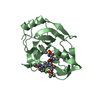
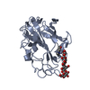
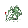
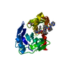
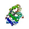
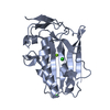
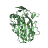
Structural insights into catalytic and substrate binding mechanisms of the strategic EndA nuclease from Streptococcus pneumoniae
Components
DNA-entry nuclease
Keywords
HYDROLASE / sequence nonspecific endonuclease
Function / homology
Function and homology information
Hydrolases; Acting on ester bonds; Endoribonucleases that are active with either ribo- or deoxyribonucleic acids and produce 5'-phosphomonoesters / establishment of competence for transformation / endonuclease activity / nucleic acid binding / hydrolase activity / metal ion binding / plasma membrane Similarity search - Function
DNA/RNA non-specific endonuclease, active site / DNA/RNA non-specific endonucleases active site. / Extracellular Endonuclease; Chain A / Extracellular Endonuclease, subunit A / DNA/RNA non-specific endonuclease / DNA/RNA non-specific endonuclease / DNA/RNA non-specific endonuclease / DNA/RNA non-specific endonuclease superfamily / 3-Layer(aba) Sandwich / Alpha Beta Similarity search - Domain/homology
Mass: 26779.693 Da / Num. of mol.: 2 / Fragment: UNP residues 31 to 274 / Mutation: H160G Source method: isolated from a genetically manipulated source Details: N-terminal His6x-GST fusion with a TEV protease cleavage site immediately preceding the EndA sequence Source: (gene. exp.) Streptococcus pneumoniae (bacteria) / Strain: TIGR4 / Gene: endA, SP_1964 / Plasmid: pET30M / Production host: Escherichia coli (E. coli) References: UniProt: P0A3S3, Hydrolases; Acting on ester bonds; Endoribonucleases that are active with either ribo- or deoxyribonucleic acids and produce 5'-phosphomonoesters
Mass: 18.015 Da / Num. of mol.: 495 / Source method: isolated from a natural source / Formula: H2O
-
Experimental details
-
Experiment
Experiment
Method: X-RAY DIFFRACTION / Number of used crystals: 1
-
Sample preparation
Crystal
Density Matthews: 2.19 Å3/Da / Density % sol: 43.72 %
Crystal grow
Temperature: 297 K / Method: vapor diffusion, sitting drop / pH: 7 Details: Crystallization solution contained 0.1M Tris pH 7, 17% PEG8000, and 0.2M MgCl2. The crystal was transferred to a hanging drop containing 0.1M Tris pH 7, 75mM MaCl, 0.2M MgCl2, and 20% ...Details: Crystallization solution contained 0.1M Tris pH 7, 17% PEG8000, and 0.2M MgCl2. The crystal was transferred to a hanging drop containing 0.1M Tris pH 7, 75mM MaCl, 0.2M MgCl2, and 20% PEG800, which was then placed over a vapor diffusion chamber containing a sitting drop bridge with 25% glutaraldehyde. The crystal was cross-linked for 8 minutes at room temperature, then transferred to a soaking solution containing 0.1M Tris pH 7, 75mM NaCl, 0.2M MgCl2,20% PEG8000, 11.6mM trimethyl lead acetate, and 9.63mM triethyl lead acetate for 43 hrs at room temperature. The soaked crystal was then transferred to a cryoprotectant (0.1M Tris pH 7, 75mM NaCl, 0.2M MgCl2, 20% PEG8000, 15% ethylene glycol, 11.9mM trimethyl lead acetate, and 13.75mM triethyl lead acetate) in four steps, then flash frozen in liquid nitrogen. , VAPOR DIFFUSION, SITTING DROP, temperature 297K
Resolution: 1.75→1.78 Å / Redundancy: 7.2 % / Mean I/σ(I) obs: 3.56 / Rsym value: 0.371 / % possible all: 68.7
-
Processing
Software
Name
Version
Classification
MAR345
datacollection
AutoBuild
modelbuilding
CNS
1.2
refinement
HKL-2000
datareduction
HKL-2000
datascaling
AutoBuild
phasing
Refinement
Method to determine structure: MOLECULAR REPLACEMENT Starting model: incomplete manually built model of EndA from a low resolution incompletely refined data set from an MBP-EndA fusion protein Resolution: 1.75→37.97 Å / Rfactor Rfree error: 0.005 / Data cutoff high absF: 91449.58 / Data cutoff low absF: 0 / Isotropic thermal model: RESTRAINED / Cross valid method: THROUGHOUT / σ(F): 0 / Stereochemistry target values: Engh & Huber / Details: BULK SOLVENT MODEL USED
In the structure databanks used in Yorodumi, some data are registered as the other names, "COVID-19 virus" and "2019-nCoV". Here are the details of the virus and the list of structure data.
Jan 31, 2019. EMDB accession codes are about to change! (news from PDBe EMDB page)
EMDB accession codes are about to change! (news from PDBe EMDB page)
The allocation of 4 digits for EMDB accession codes will soon come to an end. Whilst these codes will remain in use, new EMDB accession codes will include an additional digit and will expand incrementally as the available range of codes is exhausted. The current 4-digit format prefixed with “EMD-” (i.e. EMD-XXXX) will advance to a 5-digit format (i.e. EMD-XXXXX), and so on. It is currently estimated that the 4-digit codes will be depleted around Spring 2019, at which point the 5-digit format will come into force.
The EM Navigator/Yorodumi systems omit the EMD- prefix.
Related info.:Q: What is EMD? / ID/Accession-code notation in Yorodumi/EM Navigator
Yorodumi is a browser for structure data from EMDB, PDB, SASBDB, etc.
This page is also the successor to EM Navigator detail page, and also detail information page/front-end page for Omokage search.
The word "yorodu" (or yorozu) is an old Japanese word meaning "ten thousand". "mi" (miru) is to see.
Related info.:EMDB / PDB / SASBDB / Comparison of 3 databanks / Yorodumi Search / Aug 31, 2016. New EM Navigator & Yorodumi / Yorodumi Papers / Jmol/JSmol / Function and homology information / Changes in new EM Navigator and Yorodumi
 Movie
Movie Controller

 Yorodumi
 Open data
 Basic information
 Components
 Keywords
 Function and homology information
 Streptococcus pneumoniae (bacteria)
 X-RAY DIFFRACTION /
  Authors
 Citation
 Structure visualization
 Downloads & links
 Other downloads


 PDBj
PDBj Assembly
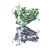

 Mass: 24.305 Da / Num. of mol.: 2 / Source method: obtained synthetically / Formula: Mg
 Mass: 35.453 Da / Num. of mol.: 5 / Source method: obtained synthetically / Formula: Cl
 Mass: 18.015 Da / Num. of mol.: 495 / Source method: isolated from a natural source / Formula: H2O
 Sample preparation
 / Beamline: 22-ID / Wavelength: 0.94 Å
 Processing