 Movie
Movie Controller

[English] 日本語
 Yorodumi
- PDB-3otv: Crystal structure of the intracellular domain of Rv3910 from Myco... -
+ Open data

- Basic information
| Entry | Database: PDB / ID: 3otv | ||||||
|---|---|---|---|---|---|---|---|
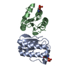
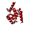
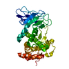
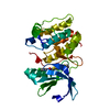
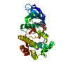
| Title | Crystal structure of the intracellular domain of Rv3910 from Mycobacterium tuberculosis | ||||||
 Components Components | PROBABLE CONSERVED TRANSMEMBRANE PROTEIN | ||||||
 Keywords Keywords | TRANSFERASE / peptidoglycan / Ser/Thr kinase / pseudokinase / Regulation / Membrane | ||||||
| Function / homology |  Function and homology information Function and homology informationlipid-linked peptidoglycan transport / lipid-linked peptidoglycan transporter activity / lipid translocation / membrane => GO:0016020 / peptidoglycan biosynthetic process / regulation of cell shape / extracellular region / plasma membrane Similarity search - Function | ||||||
| Biological species |   Mycobacterium tuberculosis (bacteria) Mycobacterium tuberculosis (bacteria) | ||||||
| Method |  X-RAY DIFFRACTION / SYNCHROTRON / MOLECULAR REPLACEMENT / Resolution: 3.094 Å X-RAY DIFFRACTION / SYNCHROTRON / MOLECULAR REPLACEMENT / Resolution: 3.094 Å | ||||||
 Authors Authors | Gee, C.L. / Alber, T. | ||||||
 Citation Citation | Journal: Sci.Signal. / Year: 2012 Title: A phosphorylated pseudokinase complex controls cell wall synthesis in mycobacteria Authors: Gee, C.L. / Papavinasasundaram, K.G. / Blair, S.R. / Baer, C.E. / Falick, A.M. / King, D.S. / Griffin, J.E. / Venghatakrishnan, H. / Zukauskas, A. / Wei, J.R. / Dhiman, R.K. / Crick, D.C. / ...Authors: Gee, C.L. / Papavinasasundaram, K.G. / Blair, S.R. / Baer, C.E. / Falick, A.M. / King, D.S. / Griffin, J.E. / Venghatakrishnan, H. / Zukauskas, A. / Wei, J.R. / Dhiman, R.K. / Crick, D.C. / Rubin, E.J. / Sassetti, C.M. / Alber, T. | ||||||
| History |
|
- Structure visualization
| Structure viewer | Molecule: MolmilJmol/JSmol |
|---|
- Downloads & links
-Download
| PDBx/mmCIF format | 3otv.cif.gz | 195.2 KB | Display | PDBx/mmCIF format |
|---|---|---|---|---|
| PDB format | pdb3otv.ent.gz | 155 KB | Display | PDB format |
| PDBx/mmJSON format | 3otv.json.gz | Tree view | PDBx/mmJSON format | |
| Others |  Other downloads Other downloads |
-Validation report
| Arichive directory | https://data.pdbj.org/pub/pdb/validation_reports/ot/3otvftp://data.pdbj.org/pub/pdb/validation_reports/ot/3otv | HTTPS FTP |
|---|
-Related structure data
| Related structure data |  3oukSC 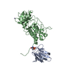 3ounC 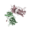 3uqcC S: Starting model for refinement C: citing same article ( |
|---|---|
| Similar structure data |


-Links
 PDBj
PDBj- Assembly
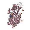
| Deposited unit | 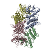
| ||||||||
|---|---|---|---|---|---|---|---|---|---|
| 1 | 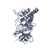
| ||||||||
| 2 | 
| ||||||||
| 3 | 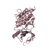
| ||||||||
| 4 | 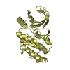
| ||||||||
| Unit cell |
|
-Components
| #1: Protein | Mass: 30133.941 Da / Num. of mol.: 4 / Fragment: Intracellular domain, UNP residues 679-963 Source method: isolated from a genetically manipulated source Source: (gene. exp.) Mycobacterium tuberculosis (bacteria) / Strain: H37Rv / Gene: MT4029, Rv3910 / Plasmid: pDEST15 / Production host: #2: Water | ChemComp-HOH / |  Mass: 18.015 Da / Num. of mol.: 60 / Source method: isolated from a natural source / Formula: H2O Mass: 18.015 Da / Num. of mol.: 60 / Source method: isolated from a natural source / Formula: H2O |
|---|
-Experimental details
-Experiment
| Experiment | Method: X-RAY DIFFRACTION / Number of used crystals: 1 |
|---|
- Sample preparation
| Crystal | Density Matthews: 2.07 Å3/Da / Density % sol: 40.52 % |
|---|---|
| Crystal grow | Temperature: 291 K / Method: vapor diffusion, hanging drop / pH: 7 Details: 0.7-0.8M succinate, pH 7.0, VAPOR DIFFUSION, HANGING DROP, temperature 291K |
-Data collection
| Diffraction | Mean temperature: 100 K |
|---|---|
| Diffraction source | Source: SYNCHROTRON / Site: ALS  / Beamline: 8.3.1 / Wavelength: 1.115872 Å / Beamline: 8.3.1 / Wavelength: 1.115872 Å |
| Detector | Type: ADSC QUANTUM 315r / Detector: CCD / Date: Sep 9, 2007 / Details: Double flat crystal, Si(111) |
| Radiation | Monochromator: Double flat crystal, Si(111) / Protocol: SINGLE WAVELENGTH / Monochromatic (M) / Laue (L): M / Scattering type: x-ray |
| Radiation wavelength | Wavelength: 1.115872 Å / Relative weight: 1 |
| Reflection | Resolution: 3.094→40.814 Å / Num. all: 18293 / Num. obs: 15986 / % possible obs: 87.4 % / Redundancy: 3.4 % / Biso Wilson estimate: 53.43 Å2 / Rmerge(I) obs: 0.631 / Rsym value: 0.113 / Net I/σ(I): 9.9 |
| Reflection shell | Resolution: 3.1→3.21 Å / Redundancy: 2.4 % / Mean I/σ(I) obs: 1.73 / Num. unique all: 1290 / Rsym value: 0.39 / % possible all: 70.8 |
- Processing
| Software |
| |||||||||||||||||||||||||||||||||||||||||||||||||
|---|---|---|---|---|---|---|---|---|---|---|---|---|---|---|---|---|---|---|---|---|---|---|---|---|---|---|---|---|---|---|---|---|---|---|---|---|---|---|---|---|---|---|---|---|---|---|---|---|---|---|
| Refinement | Method to determine structure: MOLECULAR REPLACEMENT Starting model: PDB ENTRY 3OUK Resolution: 3.094→40.814 Å / SU ML: 0.43 / Isotropic thermal model: Isotropic / Cross valid method: THROUGHOUT / σ(F): 1.33 / Stereochemistry target values: ML / Details: Phenix
| |||||||||||||||||||||||||||||||||||||||||||||||||
| Solvent computation | Shrinkage radii: 0.9 Å / VDW probe radii: 1.11 Å / Solvent model: FLAT BULK SOLVENT MODEL / Bsol: 38.537 Å2 / ksol: 0.33 e/Å3 | |||||||||||||||||||||||||||||||||||||||||||||||||
| Displacement parameters | Biso mean: 60.5 Å2
| |||||||||||||||||||||||||||||||||||||||||||||||||
| Refine analyze | Luzzati coordinate error obs: 0.512 Å | |||||||||||||||||||||||||||||||||||||||||||||||||
| Refinement step | Cycle: LAST / Resolution: 3.094→40.814 Å
| |||||||||||||||||||||||||||||||||||||||||||||||||
| Refine LS restraints |
| |||||||||||||||||||||||||||||||||||||||||||||||||
| LS refinement shell | Refine-ID: X-RAY DIFFRACTION / Total num. of bins used: 6
|
