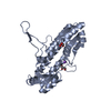
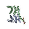
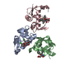
- PDB-3oss: The crystal structure of enterotoxigenic Escherichia coli GspC-Gs... -
+
Open data
ID or keywords:
Loading...
-
Basic information
Entry
Database: PDB / ID: 3oss
Title
The crystal structure of enterotoxigenic Escherichia coli GspC-GspD complex from the type II secretion system
Components
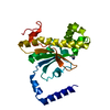
TYPE 2 SECRETION SYSTEM, GSPC
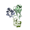
TYPE 2 SECRETION SYSTEM, SECRETIN GSPD
Keywords
PROTEIN TRANSPORT / GENERAL SECRETORY PATHWAY / HR DOMAIN / SECRETIN / LANTHANIDE-BINDING TAG
Function / homology
Function and homology information
protein secretion by the type II secretion system / type II protein secretion system complex / Secretion of toxins / cell outer membrane / identical protein binding / plasma membrane Similarity search - Function
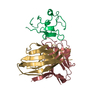
SH3 type barrels. - #830 / Type II secretion system protein GspC / Bacterial type II secretion system protein C signature. / Type II secretion system protein GspC, N-terminal / Type II secretion system protein C / Ribosomal Protein S8; Chain: A, domain 1 - #120 / Type II secretion system protein GspD / : / GspD-like, N0 domain / GspD/PilQ family ...SH3 type barrels. - #830 / Type II secretion system protein GspC / Bacterial type II secretion system protein C signature. / Type II secretion system protein GspC, N-terminal / Type II secretion system protein C / Ribosomal Protein S8; Chain: A, domain 1 - #120 / Type II secretion system protein GspD / : / GspD-like, N0 domain / GspD/PilQ family / : / NolW-like / NolW-like superfamily / Bacterial type II/III secretion system short domain / Type II/III secretion system / Bacterial type II and III secretion system protein / Ribosomal Protein S8; Chain: A, domain 1 / PDZ superfamily / SH3 type barrels. / Roll / 2-Layer Sandwich / Mainly Beta / Alpha Beta Similarity search - Domain/homology
Mass: 18.015 Da / Num. of mol.: 45 / Source method: isolated from a natural source / Formula: H2O
Sequence details
A LANTHANIDE BINDING TAG (LBT) WITH THE SEQUENCE YIDTNNDGYIEGDEL WAS INSERTED BETWEEN RESIDUES ...A LANTHANIDE BINDING TAG (LBT) WITH THE SEQUENCE YIDTNNDGYIEGDEL WAS INSERTED BETWEEN RESIDUES MET100 AND VAL113 OF GSPD
-
Experimental details
-
Experiment
Experiment
Method: X-RAY DIFFRACTION / Number of used crystals: 1
-
Sample preparation
Crystal
Density Matthews: 2.74 Å3/Da / Density % sol: 55.17 %
Crystal grow
Temperature: 294 K / Method: vapor diffusion, sitting drop / pH: 7 Details: 0.9M MG SULFATE, 0.1M BIS-TRIS PROPANE, pH 7.0, VAPOR DIFFUSION, SITTING DROP, temperature 294K
Resolution: 2.63→42.88 Å / Cor.coef. Fo:Fc: 0.934 / Cor.coef. Fo:Fc free: 0.901 / Occupancy max: 1 / Occupancy min: 1 / SU B: 29.319 / SU ML: 0.284 / SU R Cruickshank DPI: 0.5582 / Cross valid method: THROUGHOUT / ESU R: 0.558 / ESU R Free: 0.316 / Stereochemistry target values: MAXIMUM LIKELIHOOD Details: HYDROGENS HAVE BEEN ADDED IN THE RIDING POSITIONS U VALUES: WITH TLS ADDED
Rfactor
Num. reflection
% reflection
Selection details
Rfree
0.26486
457
4.9 %
RANDOM
Rwork
0.21261
-
-
-
obs
0.2151
8942
99.93 %
-
all
-
9405
-
-
Solvent computation
Ion probe radii: 0.8 Å / Shrinkage radii: 0.8 Å / VDW probe radii: 1.4 Å / Solvent model: MASK
Displacement parameters
Biso mean: 31.234 Å2
Baniso -1
Baniso -2
Baniso -3
1-
6.37 Å2
0 Å2
0 Å2
2-
-
-3.24 Å2
0 Å2
3-
-
-
-3.13 Å2
Refinement step
Cycle: LAST / Resolution: 2.63→42.88 Å
Protein
Nucleic acid
Ligand
Solvent
Total
Num. atoms
1728
0
2
45
1775
Refine LS restraints
Refine-ID
Type
Dev ideal
Dev ideal target
Number
X-RAY DIFFRACTION
r_bond_refined_d
0.013
0.022
1752
X-RAY DIFFRACTION
r_bond_other_d
0.001
0.02
1173
X-RAY DIFFRACTION
r_angle_refined_deg
1.314
1.972
2364
X-RAY DIFFRACTION
r_angle_other_deg
0.811
3
2865
X-RAY DIFFRACTION
r_dihedral_angle_1_deg
5.803
5
219
X-RAY DIFFRACTION
r_dihedral_angle_2_deg
33.865
24.39
82
X-RAY DIFFRACTION
r_dihedral_angle_3_deg
15.252
15
314
X-RAY DIFFRACTION
r_dihedral_angle_4_deg
11.148
15
15
X-RAY DIFFRACTION
r_chiral_restr
0.068
0.2
278
X-RAY DIFFRACTION
r_gen_planes_refined
0.005
0.02
1944
X-RAY DIFFRACTION
r_gen_planes_other
0.001
0.02
335
X-RAY DIFFRACTION
r_nbd_refined
X-RAY DIFFRACTION
r_nbd_other
X-RAY DIFFRACTION
r_nbtor_refined
X-RAY DIFFRACTION
r_nbtor_other
X-RAY DIFFRACTION
r_xyhbond_nbd_refined
X-RAY DIFFRACTION
r_xyhbond_nbd_other
X-RAY DIFFRACTION
r_metal_ion_refined
X-RAY DIFFRACTION
r_metal_ion_other
X-RAY DIFFRACTION
r_symmetry_vdw_refined
X-RAY DIFFRACTION
r_symmetry_vdw_other
X-RAY DIFFRACTION
r_symmetry_hbond_refined
X-RAY DIFFRACTION
r_symmetry_hbond_other
X-RAY DIFFRACTION
r_symmetry_metal_ion_refined
X-RAY DIFFRACTION
r_symmetry_metal_ion_other
X-RAY DIFFRACTION
r_mcbond_it
0.402
1.5
1102
X-RAY DIFFRACTION
r_mcbond_other
0.061
1.5
453
X-RAY DIFFRACTION
r_mcangle_it
0.807
2
1777
X-RAY DIFFRACTION
r_scbond_it
1.385
3
650
X-RAY DIFFRACTION
r_scangle_it
2.513
4.5
587
X-RAY DIFFRACTION
r_rigid_bond_restr
X-RAY DIFFRACTION
r_sphericity_free
X-RAY DIFFRACTION
r_sphericity_bonded
LS refinement shell
Resolution: 2.63→2.698 Å / Total num. of bins used: 20
Rfactor
Num. reflection
% reflection
Rfree
0.402
39
-
Rwork
0.371
650
-
obs
-
-
99.71 %
Refinement TLS params.
Method: refined / Refine-ID: X-RAY DIFFRACTION
ID
L11 (°2)
L12 (°2)
L13 (°2)
L22 (°2)
L23 (°2)
L33 (°2)
S11 (Å °)
S12 (Å °)
S13 (Å °)
S21 (Å °)
S22 (Å °)
S23 (Å °)
S31 (Å °)
S32 (Å °)
S33 (Å °)
T11 (Å2)
T12 (Å2)
T13 (Å2)
T22 (Å2)
T23 (Å2)
T33 (Å2)
Origin x (Å)
Origin y (Å)
Origin z (Å)
1
4.5248
0.895
-1.128
11.9253
-2.9487
6.7919
0.0599
-0.3147
-0.4599
0.8064
0.0524
-0.0658
0.0048
-0.0183
-0.1123
0.1654
0.0031
-0.0727
0.1288
-0.003
0.1034
-0.5426
-19.9875
28.4709
2
6.8326
-0.5278
1.4915
5.1312
-3.0885
8.052
0.276
-0.7453
-0.3235
0.9459
-0.1608
-0.4072
0.4767
0.31
-0.1152
0.382
-0.0266
-0.0705
0.1686
0.0107
0.2726
6.9874
-20.5689
30.7549
3
0.8439
-1.5798
-0.2601
5.866
1.4
4.9055
0.0605
0.0276
-0.0685
0.2459
-0.0935
0.3205
-0.1491
-0.2839
0.033
0.136
-0.0053
-0.0338
0.0825
-0.0014
0.0618
-9.0752
-7.284
20.8481
4
3.4443
0.5949
0.3839
5.76
-1.9868
8.0758
-0.0342
0.2433
0.0623
0.2458
-0.0471
-0.056
-0.0449
0.087
0.0814
0.0599
0.0203
-0.0189
0.0489
-0.0276
0.0954
-8.4647
-7.3998
-0.1067
5
1.3332
3.1544
-2.0376
31.6126
-11.2215
4.8168
-0.0086
-0.2978
0.0863
-0.0214
-0.177
-1.0859
0.032
0.341
0.1856
0.3432
0.0071
-0.0377
0.3089
-0.0746
0.1745
-5.1294
12.7875
29.7348
Refinement TLS group
ID
Refine-ID
Refine TLS-ID
Auth asym-ID
Auth seq-ID
1
X-RAY DIFFRACTION
1
C
122 - 150
2
X-RAY DIFFRACTION
2
C
151 - 186
3
X-RAY DIFFRACTION
3
D
3 - 70
4
X-RAY DIFFRACTION
3
D
74 - 80
5
X-RAY DIFFRACTION
4
D
99 - 165
6
X-RAY DIFFRACTION
5
D
699 - 800
+
About Yorodumi
-
News
-
Feb 9, 2022. New format data for meta-information of EMDB entries
New format data for meta-information of EMDB entries
Version 3 of the EMDB header file is now the official format.
The previous official version 1.9 will be removed from the archive.
In the structure databanks used in Yorodumi, some data are registered as the other names, "COVID-19 virus" and "2019-nCoV". Here are the details of the virus and the list of structure data.
Jan 31, 2019. EMDB accession codes are about to change! (news from PDBe EMDB page)
EMDB accession codes are about to change! (news from PDBe EMDB page)
The allocation of 4 digits for EMDB accession codes will soon come to an end. Whilst these codes will remain in use, new EMDB accession codes will include an additional digit and will expand incrementally as the available range of codes is exhausted. The current 4-digit format prefixed with “EMD-” (i.e. EMD-XXXX) will advance to a 5-digit format (i.e. EMD-XXXXX), and so on. It is currently estimated that the 4-digit codes will be depleted around Spring 2019, at which point the 5-digit format will come into force.
The EM Navigator/Yorodumi systems omit the EMD- prefix.
Related info.:Q: What is EMD? / ID/Accession-code notation in Yorodumi/EM Navigator
Yorodumi is a browser for structure data from EMDB, PDB, SASBDB, etc.
This page is also the successor to EM Navigator detail page, and also detail information page/front-end page for Omokage search.
The word "yorodu" (or yorozu) is an old Japanese word meaning "ten thousand". "mi" (miru) is to see.
Related info.:EMDB / PDB / SASBDB / Comparison of 3 databanks / Yorodumi Search / Aug 31, 2016. New EM Navigator & Yorodumi / Yorodumi Papers / Jmol/JSmol / Function and homology information / Changes in new EM Navigator and Yorodumi
 Movie
Movie Controller

 Yorodumi
 Open data
 Basic information
 Components
 Keywords
 Function and homology information
 X-RAY DIFFRACTION /
  Authors
 Citation
 Structure visualization
 Downloads & links
 Other downloads
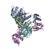


 PDBj
PDBj
 Assembly
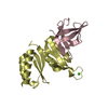

 Mass: 40.078 Da / Num. of mol.: 1 / Source method: obtained synthetically / Formula: Ca
 Mass: 35.453 Da / Num. of mol.: 1 / Source method: obtained synthetically / Formula: Cl
 Mass: 18.015 Da / Num. of mol.: 45 / Source method: isolated from a natural source / Formula: H2O
 Sample preparation
 / Beamline: BL9-2 / Wavelength: 0.97946 Å
 Processing