 Movie
Movie Controller


+ Open data

- Basic information
| Entry | Database: PDB / ID: 3orh | |||||||||
|---|---|---|---|---|---|---|---|---|---|---|
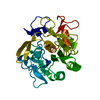
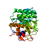
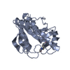
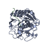
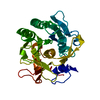
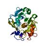
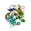
| Title | Human guanidinoacetate N-methyltransferase with SAH | |||||||||
 Components Components | Guanidinoacetate N-methyltransferase | |||||||||
 Keywords Keywords | TRANSFERASE / GUANIDINOACETATE N-METHYLTRANSFERASE / Structural Genomics / Structural Genomics Consortium / SGC | |||||||||
| Function / homology |  Function and homology information Function and homology informationguanidinoacetate N-methyltransferase / guanidinoacetate N-methyltransferase activity / creatine metabolic process / creatine biosynthetic process / Creatine metabolism / Transcriptional Regulation by MECP2 / regulation of multicellular organism growth / muscle contraction / animal organ morphogenesis / methyltransferase activity ...guanidinoacetate N-methyltransferase / guanidinoacetate N-methyltransferase activity / creatine metabolic process / creatine biosynthetic process / Creatine metabolism / Transcriptional Regulation by MECP2 / regulation of multicellular organism growth / muscle contraction / animal organ morphogenesis / methyltransferase activity / methylation / spermatogenesis / nucleus / cytosol / cytoplasm Similarity search - Function | |||||||||
| Biological species |  Homo sapiens (human) Homo sapiens (human) | |||||||||
| Method |  X-RAY DIFFRACTION / SYNCHROTRON / MOLECULAR REPLACEMENT / Resolution: 1.86 Å X-RAY DIFFRACTION / SYNCHROTRON / MOLECULAR REPLACEMENT / Resolution: 1.86 Å | |||||||||
 Authors Authors | Dong, A. / Wu, H. / Zeng, H. / Loppnau, P. / Sundstrom, M. / Arrowsmith, C.H. / Edwards, A.M. / Bochkarev, A. / Plotnikov, A.N. / Structural Genomics Consortium (SGC) | |||||||||
 Citation Citation | Journal: TO BE PUBLISHED Title: The crystal structure of human guanidinoacetate N-methyltransferase with SAH Authors: Wu, H. / Dong, A. / Zeng, H. / Loppnau, P. / Sundstrom, M. / Arrowsmith, C.H. / Edwards, A.M. / Bochkarev, A. / Plotnikov, A.N. | |||||||||
| History |
|
- Structure visualization
| Structure viewer | Molecule: MolmilJmol/JSmol |
|---|
- Downloads & links
-Download
| PDBx/mmCIF format | 3orh.cif.gz | 214.1 KB | Display | PDBx/mmCIF format |
|---|---|---|---|---|
| PDB format | pdb3orh.ent.gz | 169.5 KB | Display | PDB format |
| PDBx/mmJSON format | 3orh.json.gz | Tree view | PDBx/mmJSON format | |
| Others |  Other downloads Other downloads |
-Validation report
| Arichive directory | https://data.pdbj.org/pub/pdb/validation_reports/or/3orhftp://data.pdbj.org/pub/pdb/validation_reports/or/3orh | HTTPS FTP |
|---|
-Related structure data
| Related structure data | 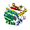 1xclS S: Starting model for refinement |
|---|---|
| Similar structure data |


-Links
 PDBj
PDBj

- Assembly
| Deposited unit | 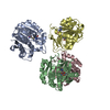
| ||||||||
|---|---|---|---|---|---|---|---|---|---|
| 1 | 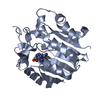
| ||||||||
| 2 | 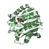
| ||||||||
| 3 | 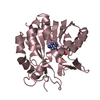
| ||||||||
| 4 | 
| ||||||||
| Unit cell |
| ||||||||
| Details | AUTHORS STATE THAT THE BIOLOGICAL ASSEMBLY IS UNKNOWN. |
-Components
| #1: Protein | Mass: 26347.123 Da / Num. of mol.: 4 Source method: isolated from a genetically manipulated source Source: (gene. exp.) Homo sapiens (human) / Gene: GAMT / Plasmid: PET28 / Production host:  References: UniProt: Q14353, guanidinoacetate N-methyltransferase #2: Chemical | ChemComp-SAH /   Type: L-peptide linking / Mass: 384.411 Da / Num. of mol.: 4 / Source method: obtained synthetically / Formula: C14H20N6O5S Type: L-peptide linking / Mass: 384.411 Da / Num. of mol.: 4 / Source method: obtained synthetically / Formula: C14H20N6O5S#3: Water | ChemComp-HOH / |  Mass: 18.015 Da / Num. of mol.: 981 / Source method: isolated from a natural source / Formula: H2O Mass: 18.015 Da / Num. of mol.: 981 / Source method: isolated from a natural source / Formula: H2O |
|---|
-Experimental details
-Experiment
| Experiment | Method: X-RAY DIFFRACTION / Number of used crystals: 1 |
|---|
- Sample preparation
| Crystal | Density Matthews: 2.86 Å3/Da / Density % sol: 57.01 % |
|---|---|
| Crystal grow | Temperature: 297 K / Method: vapor diffusion / pH: 6.5 Details: 21% PEG3350, 0.1M CACL2, 0.1M TRIS, 1MM GUANIDINE HCL, pH 6.5, VAPOR DIFFUSION, temperature 297K |
-Data collection
| Diffraction | Mean temperature: 100 K |
|---|---|
| Diffraction source | Source: SYNCHROTRON / Site: CHESS  / Beamline: A1 / Wavelength: 0.9766 Å / Beamline: A1 / Wavelength: 0.9766 Å |
| Detector | Type: ADSC QUANTUM 4 / Detector: CCD / Date: May 9, 2005 / Details: SI(111) |
| Radiation | Protocol: SINGLE WAVELENGTH / Monochromatic (M) / Laue (L): M / Scattering type: x-ray |
| Radiation wavelength | Wavelength: 0.9766 Å / Relative weight: 1 |
| Reflection | Resolution: 1.86→37.2 Å / Num. all: 97258 / Num. obs: 97258 / % possible obs: 96.5 % / Observed criterion σ(F): 0 / Observed criterion σ(I): 0 / Redundancy: 7.7 % / Biso Wilson estimate: 21.8 Å2 / Rmerge(I) obs: 0.7 / Rsym value: 0.7 / Net I/σ(I): 21.2 |
| Reflection shell | Resolution: 1.86→1.89 Å / Redundancy: 4.1 % / Rmerge(I) obs: 0.407 / Mean I/σ(I) obs: 2.3 / Num. unique all: 3582 / % possible all: 74 |
- Processing
| Software |
| ||||||||||||||||||||||||||||||||||||||||||||||||||||||||||||||||||||||||||||||||||||||||||
|---|---|---|---|---|---|---|---|---|---|---|---|---|---|---|---|---|---|---|---|---|---|---|---|---|---|---|---|---|---|---|---|---|---|---|---|---|---|---|---|---|---|---|---|---|---|---|---|---|---|---|---|---|---|---|---|---|---|---|---|---|---|---|---|---|---|---|---|---|---|---|---|---|---|---|---|---|---|---|---|---|---|---|---|---|---|---|---|---|---|---|---|
| Refinement | Method to determine structure: MOLECULAR REPLACEMENT Starting model: PDB entry 1XCL Resolution: 1.86→37.2 Å / Cor.coef. Fo:Fc: 0.9154 / Cor.coef. Fo:Fc free: 0.8976 / Occupancy max: 1 / Occupancy min: 0.5 / Cross valid method: THROUGHOUT / σ(F): 0 / σ(I): 0
| ||||||||||||||||||||||||||||||||||||||||||||||||||||||||||||||||||||||||||||||||||||||||||
| Displacement parameters | Biso max: 207.74 Å2 / Biso mean: 24.6722 Å2 / Biso min: 8.44 Å2
| ||||||||||||||||||||||||||||||||||||||||||||||||||||||||||||||||||||||||||||||||||||||||||
| Refine analyze | Luzzati coordinate error obs: 0.234 Å | ||||||||||||||||||||||||||||||||||||||||||||||||||||||||||||||||||||||||||||||||||||||||||
| Refinement step | Cycle: LAST / Resolution: 1.86→37.2 Å
| ||||||||||||||||||||||||||||||||||||||||||||||||||||||||||||||||||||||||||||||||||||||||||
| Refine LS restraints |
| ||||||||||||||||||||||||||||||||||||||||||||||||||||||||||||||||||||||||||||||||||||||||||
| LS refinement shell | Resolution: 1.86→1.91 Å / Total num. of bins used: 20
|
