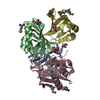
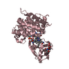
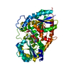
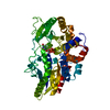
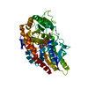
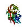
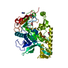
- PDB-3oqq: Crystal structure of a Putative lipoprotein (BACOVA_00967) from B... -
+
Open data
ID or keywords:
Loading...
-
Basic information
Entry
Database: PDB / ID: 3oqq
Title
Crystal structure of a Putative lipoprotein (BACOVA_00967) from Bacteroides ovatus at 2.08 A resolution
Components
Putative lipoprotein
Keywords
Structural Genomics / Unknown Function / EXTRACELLULAR PROTEIN / JOINT CENTER FOR STRUCTURAL GENOMICS / JCSG / PROTEIN STRUCTURE INITIATIVE / PSI-2
Function / homology
LruC domain / Domain of unknown function DUF4841 / Domain of unknown function DUF4842 / Domain of unknown function (DUF4841) / Domain of unknown function (DUF4842) / metal ion binding / ACETATE ION / TRIETHYLENE GLYCOL / Uncharacterized protein
Function and homology information
Biological species
Bacteroides ovatus (bacteria)
Method
X-RAY DIFFRACTION / SYNCHROTRON / MAD / Resolution: 2.08 Å
ANALYTICAL SIZE EXCLUSION CHROMATOGRAPHY WITH STATIC LIGHT SCATTERING SUPPORTS THE ASSIGNMENT OF A MONOMER AS A SIGNIFICANT OLIGOMERIZATION STATE IN SOLUTION.
-
Components
#1: Protein
Putativelipoprotein
Mass: 48892.066 Da / Num. of mol.: 1 Source method: isolated from a genetically manipulated source Source: (gene. exp.) Bacteroides ovatus (bacteria) / Strain: ATCC 8483 / Gene: BACOVA_00967 / Plasmid: SpeedET / Production host: Escherichia Coli (E. coli) / Strain (production host): HK100 / References: UniProt: A7LT28
Mass: 18.015 Da / Num. of mol.: 301 / Source method: isolated from a natural source / Formula: H2O
Has protein modification
Y
Sequence details
THE CONSTRUCT (RESIDUES 23-456) WAS EXPRESSED WITH A PURIFICATION TAG MGSDKIHHHHHHENLYFQG. THE TAG ...THE CONSTRUCT (RESIDUES 23-456) WAS EXPRESSED WITH A PURIFICATION TAG MGSDKIHHHHHHENLYFQG. THE TAG WAS REMOVED WITH TEV PROTEASE LEAVING ONLY A GLYCINE (0) FOLLOWED BY THE TARGET SEQUENCE.
-
Experimental details
-
Experiment
Experiment
Method: X-RAY DIFFRACTION / Number of used crystals: 2
-
Sample preparation
Crystal
ID
Density Matthews (Å3/Da)
Density % sol (%)
Description
1
2.48
50.5
THREE-WAVELENGTH MAD PHASES WERE OBTAINED FROM TWO CRYSTALS. THE PEAK DATA WERE FROM ONE CRYSTAL DIFFRACTING TO 2.08 A. THE INFLECTION AND REMOTE DATA WERE FROM ANOTHER CRYSTAL DIFFRACTING TO 2.6 A. THE PEAK DATA (FROM THE HIGHER RESOLUTION CRYSTAL) WERE USED FOR REFINEMENT.
2
Crystal grow
Temperature (K)
Crystal-ID
Method
pH
Details
277
1
vapor diffusion, sitting drop
8
0.2M Ca(OAc)2, 20.0% PEG-1000, 0.1M Imidazole pH 8.0, VAPOR DIFFUSION,SITTING DROP,NANODROP, temperature 277K, VAPOR DIFFUSION, SITTING DROP
277
2
vapor diffusion, sitting drop
8
0.2M Ca(OAc)2, 20.0% PEG-1000, 0.1M Imidazole pH 8.0, VAPOR DIFFUSION,SITTING DROP,NANODROP, temperature 277K, VAPOR DIFFUSION, SITTING DROP
-
Data collection
Diffraction
ID
Mean temperature (K)
Crystal-ID
1
100
1
2
100
2
Diffraction source
Source
Site
Beamline
ID
Wavelength (Å)
SYNCHROTRON
SSRL
BL9-2
1
0.97903
SYNCHROTRON
SSRL
BL9-2
2
0.97918,0.91837
Detector
Type
ID
Detector
Date
Details
MARMOSAIC 325 mm CCD
1
CCD
Jul 22, 2010
Flatcollimatingmirror, toroidfocusingmirror
MARMOSAIC 325 mm CCD
2
CCD
Jul 22, 2010
Flatcollimatingmirror, toroidfocusingmirror
Radiation
ID
Monochromator
Protocol
Monochromatic (M) / Laue (L)
Scattering type
Wavelength-ID
1
Doublecrystalmonochromator
MAD
M
x-ray
1
2
Doublecrystalmonochromator
MAD
M
x-ray
2
Radiation wavelength
ID
Wavelength (Å)
Relative weight
1
0.97903
1
2
0.97918
1
3
0.91837
1
Reflection
Resolution: 2.08→44.623 Å / Num. obs: 28606 / % possible obs: 99.5 % / Observed criterion σ(I): -3 / Redundancy: 3.8 % / Biso Wilson estimate: 27.02 Å2 / Rmerge(I) obs: 0.109 / Net I/σ(I): 9.31
Reflection shell
Diffraction-ID: 1,2
Resolution (Å)
Redundancy (%)
Rmerge(I) obs
Mean I/σ(I) obs
Num. measured obs
Num. unique obs
% possible all
2.08-2.15
3.7
0.567
2.1
10236
2722
99.9
2.15-2.24
3.8
0.441
2.7
11288
2995
99.5
2.24-2.34
3.8
0.382
3.2
10326
2772
99.5
2.34-2.47
3.8
0.324
3.8
11427
3016
99.5
2.47-2.62
3.8
0.264
4.6
10395
2749
100
2.62-2.82
3.8
0.197
6.1
10711
2821
99.6
2.82-3.1
3.8
0.13
9
10811
2847
99.6
3.1-3.55
3.7
0.071
14.9
10893
2884
99.4
3.55-4.47
3.8
0.048
21.2
10788
2874
99.2
4.47-44.6
3.8
0.046
25
10884
2921
99.3
-
Phasing
Phasing
Method: MAD
-
Processing
Software
Name
Version
Classification
NB
SHELX
phasing
BUSTER-TNT
BUSTER2.8.0
refinement
XSCALE
dataprocessing
PDB_EXTRACT
3.1
dataextraction
XDS
datareduction
XSCALE
datascaling
SHELXD
phasing
autoSHARP
phasing
BUSTER
2.8.0
refinement
Refinement
Method to determine structure: MAD / Resolution: 2.08→25.75 Å / Cor.coef. Fo:Fc: 0.9493 / Cor.coef. Fo:Fc free: 0.9287 / Occupancy max: 1 / Occupancy min: 0.5 / Cross valid method: THROUGHOUT / σ(F): 0 Details: 1. A MET-INHIBITION PROTOCOL WAS USED FOR SELENOMETHIONINE INCORPORATION DURING PROTEIN EXPRESSION. THE OCCUPANCY OF THE SE ATOMS IN THE MSE RESIDUES WAS REDUCED TO 0.75 FOR THE REDUCED ...Details: 1. A MET-INHIBITION PROTOCOL WAS USED FOR SELENOMETHIONINE INCORPORATION DURING PROTEIN EXPRESSION. THE OCCUPANCY OF THE SE ATOMS IN THE MSE RESIDUES WAS REDUCED TO 0.75 FOR THE REDUCED SCATTERING POWER DUE TO PARTIAL S-MET INCORPORATION. 2. ATOM RECORD CONTAINS SUM OF TLS AND RESIDUAL B FACTORS. ANISOU RECORD CONTAINS SUM OF TLS AND RESIDUAL U FACTORS. 3. CALCIUM (CA), ACETATE, POLYETHYLENE GLYCOL FRAGMENTS (PGE) MODELED ARE PRESENT PROTEIN/CRYSTALLIZATION/CRYO BUFFER. 4. RAMACHANDRAN OUTLIERS (A66 AND A210) ARE SUPPORTED BY DENSITY. 5. THE MAD PHASES WERE USED AS RESTRAINTS DURING REFINEMENT.
Rfactor
Num. reflection
% reflection
Selection details
Rfree
0.2039
1449
5.07 %
RANDOM
Rwork
0.161
-
-
-
obs
0.1633
28586
-
-
Displacement parameters
Biso mean: 31.81 Å2
Baniso -1
Baniso -2
Baniso -3
1-
5.1771 Å2
0 Å2
-3.8575 Å2
2-
-
-3.7928 Å2
0 Å2
3-
-
-
-1.3843 Å2
Refinement step
Cycle: LAST / Resolution: 2.08→25.75 Å
Protein
Nucleic acid
Ligand
Solvent
Total
Num. atoms
3223
0
25
301
3549
Refine LS restraints
Refine-ID
Type
Dev ideal
Number
Restraint function
Weight
X-RAY DIFFRACTION
t_bond_d
0.01
3328
HARMONIC
2
X-RAY DIFFRACTION
t_angle_deg
1.08
4517
HARMONIC
2
X-RAY DIFFRACTION
t_dihedral_angle_d
1539
SINUSOIDAL
2
X-RAY DIFFRACTION
t_incorr_chiral_ct
X-RAY DIFFRACTION
t_pseud_angle
X-RAY DIFFRACTION
t_trig_c_planes
95
HARMONIC
2
X-RAY DIFFRACTION
t_gen_planes
478
HARMONIC
5
X-RAY DIFFRACTION
t_it
3328
HARMONIC
20
X-RAY DIFFRACTION
t_nbd
0
SEMIHARMONIC
5
X-RAY DIFFRACTION
t_omega_torsion
3.93
X-RAY DIFFRACTION
t_other_torsion
2.91
X-RAY DIFFRACTION
t_improper_torsion
X-RAY DIFFRACTION
t_chiral_improper_torsion
431
SEMIHARMONIC
5
X-RAY DIFFRACTION
t_sum_occupancies
X-RAY DIFFRACTION
t_utility_distance
X-RAY DIFFRACTION
t_utility_angle
X-RAY DIFFRACTION
t_utility_torsion
X-RAY DIFFRACTION
t_ideal_dist_contact
4072
SEMIHARMONIC
4
LS refinement shell
Resolution: 2.08→2.16 Å / Total num. of bins used: 14
Rfactor
Num. reflection
% reflection
Rfree
0.2556
130
4.32 %
Rwork
0.1848
2881
-
all
0.1876
3011
-
Refinement TLS params.
Method: refined / Origin x: 11.7092 Å / Origin y: -0.349 Å / Origin z: 25.9619 Å
11
12
13
21
22
23
31
32
33
T
-0.1042 Å2
0.0044 Å2
0.0103 Å2
-
0.015 Å2
0.005 Å2
-
-
-0.0997 Å2
L
1.4399 °2
0.2403 °2
-0.0677 °2
-
0.4378 °2
0.0674 °2
-
-
0.3739 °2
S
0.0192 Å °
-0.1222 Å °
-0.0968 Å °
0.0286 Å °
-0.0233 Å °
-0.0755 Å °
0.0376 Å °
0.0136 Å °
0.0042 Å °
Refinement TLS group
Selection details: { A| 40 - 456 }
+
About Yorodumi
-
News
-
Feb 9, 2022. New format data for meta-information of EMDB entries
New format data for meta-information of EMDB entries
Version 3 of the EMDB header file is now the official format.
The previous official version 1.9 will be removed from the archive.
In the structure databanks used in Yorodumi, some data are registered as the other names, "COVID-19 virus" and "2019-nCoV". Here are the details of the virus and the list of structure data.
Jan 31, 2019. EMDB accession codes are about to change! (news from PDBe EMDB page)
EMDB accession codes are about to change! (news from PDBe EMDB page)
The allocation of 4 digits for EMDB accession codes will soon come to an end. Whilst these codes will remain in use, new EMDB accession codes will include an additional digit and will expand incrementally as the available range of codes is exhausted. The current 4-digit format prefixed with “EMD-” (i.e. EMD-XXXX) will advance to a 5-digit format (i.e. EMD-XXXXX), and so on. It is currently estimated that the 4-digit codes will be depleted around Spring 2019, at which point the 5-digit format will come into force.
The EM Navigator/Yorodumi systems omit the EMD- prefix.
Related info.:Q: What is EMD? / ID/Accession-code notation in Yorodumi/EM Navigator
Yorodumi is a browser for structure data from EMDB, PDB, SASBDB, etc.
This page is also the successor to EM Navigator detail page, and also detail information page/front-end page for Omokage search.
The word "yorodu" (or yorozu) is an old Japanese word meaning "ten thousand". "mi" (miru) is to see.
Related info.:EMDB / PDB / SASBDB / Comparison of 3 databanks / Yorodumi Search / Aug 31, 2016. New EM Navigator & Yorodumi / Yorodumi Papers / Jmol/JSmol / Function and homology information / Changes in new EM Navigator and Yorodumi
 Movie
Movie Controller

 Yorodumi
 Open data
 Basic information
 Components
 Keywords
 Function and homology information
 Bacteroides ovatus (bacteria)
 X-RAY DIFFRACTION /
  Authors
 Citation
 Structure visualization
 Downloads & links
 Other downloads
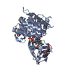

 PDBj
PDBj Assembly
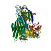

 Mass: 40.078 Da / Num. of mol.: 4 / Source method: obtained synthetically / Formula: Ca
 Mass: 59.044 Da / Num. of mol.: 1 / Source method: obtained synthetically / Formula: C2H3O2
 Mass: 150.173 Da / Num. of mol.: 2 / Source method: obtained synthetically / Formula: C6H14O4
 Mass: 18.015 Da / Num. of mol.: 301 / Source method: isolated from a natural source / Formula: H2O
 Sample preparation
 Processing