Mass: 18.015 Da / Num. of mol.: 57 / Source method: isolated from a natural source / Formula: H2O
Has protein modification
Y
Sequence details
THE CONSTRUCT WAS EXPRESSED WITH A PURIFICATION TAG MGSDKIHHHHHHENLYFQG. THE TAG WAS REMOVED WITH ...THE CONSTRUCT WAS EXPRESSED WITH A PURIFICATION TAG MGSDKIHHHHHHENLYFQG. THE TAG WAS REMOVED WITH TEV PROTEASE LEAVING ONLY A GLYCINE (0) FOLLOWED BY THE TARGET SEQUENCE.
-
Experimental details
-
Experiment
Experiment
Method: X-RAY DIFFRACTION / Number of used crystals: 1
-
Sample preparation
Crystal
Density Matthews: 2.22 Å3/Da / Density % sol: 44.48 %
Crystal grow
Temperature: 293 K / Method: vapor diffusion, sitting drop / pH: 8.21 Details: 0.84M sodium citrate, 0.1M Imidazole pH 8.21, NANODROP, VAPOR DIFFUSION, SITTING DROP, temperature 293K
Type: MARMOSAIC 325 mm CCD / Detector: CCD / Date: Jun 10, 2010 / Details: Flat mirror (vertical focusing)
Radiation
Monochromator: Single crystal Si(111) bent monochromator (horizontal focusing) Protocol: MAD / Monochromatic (M) / Laue (L): M / Scattering type: x-ray
Radiation wavelength
ID
Wavelength (Å)
Relative weight
1
0.91837
1
2
0.97922
1
3
0.97898
1
Reflection
Resolution: 2.54→29.88 Å / Num. obs: 37971 / % possible obs: 99.3 % / Observed criterion σ(I): -3 / Biso Wilson estimate: 70.646 Å2 / Rmerge(I) obs: 0.074 / Net I/σ(I): 11.66
Reflection shell
Diffraction-ID: 1
Resolution (Å)
Highest resolution (Å)
Rmerge(I) obs
Mean I/σ(I) obs
Num. measured obs
Num. unique obs
% possible all
2.55-2.64
0.822
1.6
25491
6488
94.1
2.64-2.75
0.608
2.3
29122
7199
99.9
2.75-2.87
0.468
3
27020
6646
100
2.87-3.02
0.357
3.8
27929
6877
100
3.02-3.21
0.239
5.7
28660
7059
100
3.21-3.46
0.143
9.3
28214
6931
100
3.46-3.8
0.084
15.1
27761
6846
100
3.8-4.35
0.06
21.4
28129
6985
100
4.35-5.46
0.049
24.6
27811
6898
100
5.46
0.035
28.9
28082
7085
99.1
-
Phasing
Phasing
Method: MAD
-
Processing
Software
Name
Version
Classification
NB
SHELX
phasing
BUSTER-TNT
BUSTER2.8.0
refinement
XSCALE
dataprocessing
PDB_EXTRACT
3.1
dataextraction
XDS
datareduction
XSCALE
datascaling
SHELXD
phasing
autoSHARP
phasing
BUSTER
2.8.0
refinement
Refinement
Method to determine structure: MAD / Resolution: 2.54→29.88 Å / Cor.coef. Fo:Fc: 0.9546 / Cor.coef. Fo:Fc free: 0.9399 / Occupancy max: 1 / Occupancy min: 0.5 / Cross valid method: THROUGHOUT / σ(F): 0 Details: 1. A MET-INHIBITION PROTOCOL WAS USED FOR SELENOMETHIONINE INCORPORATION DURING PROTEIN EXPRESSION. THE OCCUPANCY OF THE SE ATOMS IN THE MSE RESIDUES WAS REDUCED TO 0.75 FOR THE REDUCED ...Details: 1. A MET-INHIBITION PROTOCOL WAS USED FOR SELENOMETHIONINE INCORPORATION DURING PROTEIN EXPRESSION. THE OCCUPANCY OF THE SE ATOMS IN THE MSE RESIDUES WAS REDUCED TO 0.75 FOR THE REDUCED SCATTERING POWER DUE TO PARTIAL S-MET INCORPORATION. 2. NCS RESTRAINTS WERE APPLIED USING BUSTER'S LSSR RESTRAINT REPRESENTATION (-AUTONCS). 3. ATOM RECORD CONTAINS SUM OF TLS AND RESIDUAL B FACTORS. ANISOU RECORD CONTAINS SUM OF TLS AND RESIDUAL U FACTORS.
In the structure databanks used in Yorodumi, some data are registered as the other names, "COVID-19 virus" and "2019-nCoV". Here are the details of the virus and the list of structure data.
Jan 31, 2019. EMDB accession codes are about to change! (news from PDBe EMDB page)
EMDB accession codes are about to change! (news from PDBe EMDB page)
The allocation of 4 digits for EMDB accession codes will soon come to an end. Whilst these codes will remain in use, new EMDB accession codes will include an additional digit and will expand incrementally as the available range of codes is exhausted. The current 4-digit format prefixed with “EMD-” (i.e. EMD-XXXX) will advance to a 5-digit format (i.e. EMD-XXXXX), and so on. It is currently estimated that the 4-digit codes will be depleted around Spring 2019, at which point the 5-digit format will come into force.
The EM Navigator/Yorodumi systems omit the EMD- prefix.
Related info.:Q: What is EMD? / ID/Accession-code notation in Yorodumi/EM Navigator
Yorodumi is a browser for structure data from EMDB, PDB, SASBDB, etc.
This page is also the successor to EM Navigator detail page, and also detail information page/front-end page for Omokage search.
The word "yorodu" (or yorozu) is an old Japanese word meaning "ten thousand". "mi" (miru) is to see.
Related info.:EMDB / PDB / SASBDB / Comparison of 3 databanks / Yorodumi Search / Aug 31, 2016. New EM Navigator & Yorodumi / Yorodumi Papers / Jmol/JSmol / Function and homology information / Changes in new EM Navigator and Yorodumi
 Movie
Movie Controller

 Yorodumi
 Open data
 Basic information
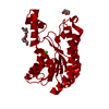
 Components
 Keywords
 Function and homology information
 Pseudomonas syringae pv. tomato (bacteria)
 X-RAY DIFFRACTION /
  Authors
 Citation
 Structure visualization
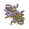
 Downloads & links
 Other downloads
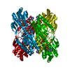
 PDBj
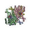
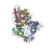
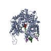
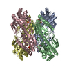
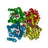
PDBj Assembly
 Mass: 18.015 Da / Num. of mol.: 57 / Source method: isolated from a natural source / Formula: H2O
 Sample preparation
 / Beamline: BL11-1 / Wavelength: 0.91837,0.97922,0.97898
 Processing