 Movie
Movie Controller

[English] 日本語
 Yorodumi
- PDB-3vdq: Crystal structure of alcaligenes faecalis D-3-hydroxybutyrate deh... -
+ Open data

- Basic information
| Entry | Database: PDB / ID: 3vdq | |||||||||
|---|---|---|---|---|---|---|---|---|---|---|
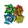
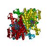
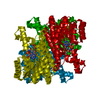
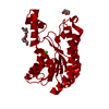
| Title | Crystal structure of alcaligenes faecalis D-3-hydroxybutyrate dehydrogenase in complex with NAD(+) and acetate | |||||||||
 Components Components | D-3-hydroxybutyrate dehydrogenase | |||||||||
 Keywords Keywords | OXIDOREDUCTASE / NAD DEPENDENT ENZYME / HYDROXYBUTYRATE DEHYDROGENASE / KETONE BODIES | |||||||||
| Function / homology |  Function and homology information Function and homology information3-hydroxybutyrate dehydrogenase / 3-hydroxybutyrate dehydrogenase activity / monocarboxylic acid metabolic process / nucleotide binding / metal ion binding Similarity search - Function | |||||||||
| Biological species |  Alcaligenes faecalis (bacteria) Alcaligenes faecalis (bacteria) | |||||||||
| Method |  X-RAY DIFFRACTION / SYNCHROTRON / MOLECULAR REPLACEMENT / Resolution: 2.2 Å X-RAY DIFFRACTION / SYNCHROTRON / MOLECULAR REPLACEMENT / Resolution: 2.2 Å | |||||||||
 Authors Authors | Hoque, M.M. / Shimizu, S. / Hossain, M.T. / Yamamoto, T. / Suzuki, K. / Takenaka, A. | |||||||||
 Citation Citation | Journal: Acta Crystallogr.,Sect.D / Year: 2008 Title: The structures of Alcaligenes faecalis D-3-hydroxybutyrate dehydrogenase before and after NAD+ and acetate binding suggest a dynamical reaction mechanism as a member of the SDR family. Authors: Hoque, M.M. / Shimizu, S. / Hossain, M.T. / Yamamoto, T. / Imamura, S. / Suzuki, K. / Tsunoda, M. / Amano, H. / Sekiguchi, T. / Takenaka, A. | |||||||||
| History |
|
- Structure visualization
| Structure viewer | Molecule: MolmilJmol/JSmol |
|---|
- Downloads & links
-Download
| PDBx/mmCIF format | 3vdq.cif.gz | 221.8 KB | Display | PDBx/mmCIF format |
|---|---|---|---|---|
| PDB format | pdb3vdq.ent.gz | 177.5 KB | Display | PDB format |
| PDBx/mmJSON format | 3vdq.json.gz | Tree view | PDBx/mmJSON format | |
| Others |  Other downloads Other downloads |
-Validation report
| Arichive directory | https://data.pdbj.org/pub/pdb/validation_reports/vd/3vdqftp://data.pdbj.org/pub/pdb/validation_reports/vd/3vdq | HTTPS FTP |
|---|
-Related structure data
| Related structure data |  2yz7SC S: Starting model for refinement C: citing same article ( |
|---|---|
| Similar structure data |


-Links
 PDBj
PDBj


- Assembly
| Deposited unit | 
| ||||||||
|---|---|---|---|---|---|---|---|---|---|
| 1 |
| ||||||||
| Unit cell |
|
-Components
-Protein , 1 types, 4 molecules ABCD
| #1: Protein | Mass: 27118.943 Da / Num. of mol.: 4 / Source method: isolated from a natural source / Source: (natural) Alcaligenes faecalis (bacteria)References: UniProt: D0VWQ0, 3-hydroxybutyrate dehydrogenase |
|---|
-Non-polymers , 5 types, 845 molecules 


| #2: Chemical | ChemComp-ACT /  Mass: 59.044 Da / Num. of mol.: 4 / Source method: obtained synthetically / Formula: C2H3O2 Mass: 59.044 Da / Num. of mol.: 4 / Source method: obtained synthetically / Formula: C2H3O2#3: Chemical | ChemComp-CL /  Mass: 35.453 Da / Num. of mol.: 4 / Source method: obtained synthetically / Formula: Cl Mass: 35.453 Da / Num. of mol.: 4 / Source method: obtained synthetically / Formula: Cl#4: Chemical | ChemComp-NAD /  Mass: 663.425 Da / Num. of mol.: 4 / Source method: obtained synthetically / Formula: C21H27N7O14P2 / Comment: NAD*YM Mass: 663.425 Da / Num. of mol.: 4 / Source method: obtained synthetically / Formula: C21H27N7O14P2 / Comment: NAD*YM#5: Chemical |  Mass: 40.078 Da / Num. of mol.: 2 / Source method: obtained synthetically / Formula: Ca Mass: 40.078 Da / Num. of mol.: 2 / Source method: obtained synthetically / Formula: Ca#6: Water | ChemComp-HOH / | Mass: 18.015 Da / Num. of mol.: 831 / Source method: isolated from a natural source / Formula: H2O |
|---|
-Experimental details
-Experiment
| Experiment | Method: X-RAY DIFFRACTION / Number of used crystals: 1 |
|---|
- Sample preparation
| Crystal | Density Matthews: 2.52 Å3/Da / Density % sol: 51.25 % |
|---|---|
| Crystal grow | Temperature: 277 K / Method: vapor diffusion, hanging drop / pH: 8.5 Details: 30% PEG 4000, 0.2M SODIUM ACETATE TRIHYDRATE, 1.0M TRIS-HCL BUFFER, pH 8.5, VAPOR DIFFUSION, HANGING DROP, temperature 277K |
-Data collection
| Diffraction | Mean temperature: 100 K |
|---|---|
| Diffraction source | Source: SYNCHROTRON / Site: Photon Factory  / Beamline: BL-17A / Wavelength: 1 Å / Beamline: BL-17A / Wavelength: 1 Å |
| Detector | Type: ADSC QUANTUM 270 / Detector: CCD / Date: Jun 17, 2007 / Details: mirrors |
| Radiation | Monochromator: FLAT SI(111) / Protocol: SINGLE WAVELENGTH / Monochromatic (M) / Laue (L): M / Scattering type: x-ray |
| Radiation wavelength | Wavelength: 1 Å / Relative weight: 1 |
| Reflection | Resolution: 2.2→46.01 Å / Num. obs: 57443 / % possible obs: 100 % / Redundancy: 12.47 % / Rmerge(I) obs: 0.133 / Net I/σ(I): 12.3 |
| Reflection shell | Resolution: 2.2→2.28 Å / Redundancy: 12.58 % / Rmerge(I) obs: 0.317 / Mean I/σ(I) obs: 6.6 / Num. unique all: 5650 / Rsym value: 0.1304 / % possible all: 100 |
- Processing
| Software |
| |||||||||||||||||||||||||
|---|---|---|---|---|---|---|---|---|---|---|---|---|---|---|---|---|---|---|---|---|---|---|---|---|---|---|
| Refinement | Method to determine structure: MOLECULAR REPLACEMENT Starting model: 2YZ7 Resolution: 2.2→40 Å / Isotropic thermal model: Anisotropic / Cross valid method: THROUGHOUT / σ(F): 0 / Stereochemistry target values: Engh & Huber
| |||||||||||||||||||||||||
| Displacement parameters |
| |||||||||||||||||||||||||
| Refine analyze | Luzzati coordinate error obs: 0.25 Å / Luzzati d res low obs: 40 Å / Luzzati sigma a obs: 0.26 Å | |||||||||||||||||||||||||
| Refinement step | Cycle: LAST / Resolution: 2.2→40 Å
|
