 Movie
Movie Controller

[English] 日本語
 Yorodumi
- PDB-3oq3: Structural Basis of Type-I Interferon Sequestration by a Poxvirus... -
+ Open data

- Basic information
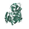
| Entry | Database: PDB / ID: 3oq3 | ||||||
|---|---|---|---|---|---|---|---|
| Title | Structural Basis of Type-I Interferon Sequestration by a Poxvirus Decoy Receptor | ||||||
 Components Components |
| ||||||
 Keywords Keywords | CYTOKINE/VIRAL PROTEIN / Ectromelia / Mousepox Virus / Moscow strain / Cytokine decoy Receptor / Virus/Viral protein / Type-1 Interferon / Soluble a/b-IFNR / Viral immune evasion / Immunoglobulin Domain / Structural Genomics / Center for Structural Genomics of Infectious Diseases / CSGID / Immunoglobulin-like / IFN-alpha/beta binding protein / IFN-alpha / extracellular / secreted / CYTOKINE-VIRAL PROTEIN complex | ||||||
| Function / homology |  Function and homology information Function and homology informationRegulation of IFNA/IFNB signaling / type I interferon receptor binding / B cell activation involved in immune response / Interferon alpha/beta signaling / natural killer cell activation involved in immune response / T cell activation involved in immune response / type I interferon-mediated signaling pathway / response to exogenous dsRNA / humoral immune response / cytokine activity ...Regulation of IFNA/IFNB signaling / type I interferon receptor binding / B cell activation involved in immune response / Interferon alpha/beta signaling / natural killer cell activation involved in immune response / T cell activation involved in immune response / type I interferon-mediated signaling pathway / response to exogenous dsRNA / humoral immune response / cytokine activity / defense response to virus / adaptive immune response / symbiont-mediated suppression of host innate immune response / symbiont-mediated suppression of host type I interferon-mediated signaling pathway / : / extracellular region / metal ion binding Similarity search - Function | ||||||
| Biological species |   Ectromelia virus Ectromelia virus | ||||||
| Method |  X-RAY DIFFRACTION / SYNCHROTRON / SAD/molecular replacement / Resolution: 2.1 Å X-RAY DIFFRACTION / SYNCHROTRON / SAD/molecular replacement / Resolution: 2.1 Å | ||||||
 Authors Authors | Fremont, D.H. / Lee, C.A. / Center for Structural Genomics of Infectious Diseases (CSGID) | ||||||
 Citation Citation | Journal: To be Published Title: Structural Basis of Type-I Interferon Sequestration by a Poxvirus Decoy Receptor Authors: Fremont, D.H. / Lee, C.A. / Center for Structural Genomics of Infectious Diseases (CSGID) | ||||||
| History |
|
- Structure visualization
| Structure viewer | Molecule: MolmilJmol/JSmol |
|---|
- Downloads & links
-Download
| PDBx/mmCIF format | 3oq3.cif.gz | 124.4 KB | Display | PDBx/mmCIF format |
|---|---|---|---|---|
| PDB format | pdb3oq3.ent.gz | 93.8 KB | Display | PDB format |
| PDBx/mmJSON format | 3oq3.json.gz | Tree view | PDBx/mmJSON format | |
| Others |  Other downloads Other downloads |
-Validation report
| Arichive directory | https://data.pdbj.org/pub/pdb/validation_reports/oq/3oq3ftp://data.pdbj.org/pub/pdb/validation_reports/oq/3oq3 | HTTPS FTP |
|---|
-Related structure data
| Related structure data |  1itfS S: Starting model for refinement |
|---|---|
| Similar structure data | |
| Other databases |


-Links
 PDBj
PDBj


- Assembly
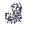
| Deposited unit | 
| ||||||||
|---|---|---|---|---|---|---|---|---|---|
| 1 |
| ||||||||
| Unit cell |
|
-Components
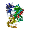
-Protein / Antibody , 2 types, 2 molecules AB
| #1: Protein | Mass: 18985.826 Da / Num. of mol.: 1 Source method: isolated from a genetically manipulated source Source: (gene. exp.)  |
|---|---|
| #2: Antibody | Mass: 37626.359 Da / Num. of mol.: 1 Source method: isolated from a genetically manipulated source Source: (gene. exp.) Ectromelia virus / Strain: Moscow / Gene: C12R, EVM166 / Plasmid: pET23B(+) / Production host: |
-Non-polymers , 6 types, 371 molecules 


| #3: Chemical | ChemComp-ZN /  Mass: 65.409 Da / Num. of mol.: 1 / Source method: obtained synthetically / Formula: Zn Mass: 65.409 Da / Num. of mol.: 1 / Source method: obtained synthetically / Formula: Zn | ||||||
|---|---|---|---|---|---|---|---|
| #4: Chemical | ChemComp-CL /  Mass: 35.453 Da / Num. of mol.: 1 / Source method: obtained synthetically / Formula: Cl Mass: 35.453 Da / Num. of mol.: 1 / Source method: obtained synthetically / Formula: Cl | ||||||
| #5: Chemical | ChemComp-EDO /  Mass: 62.068 Da / Num. of mol.: 4 / Source method: obtained synthetically / Formula: C2H6O2 Mass: 62.068 Da / Num. of mol.: 4 / Source method: obtained synthetically / Formula: C2H6O2#6: Chemical |  Mass: 59.044 Da / Num. of mol.: 2 / Source method: obtained synthetically / Formula: C2H3O2 Mass: 59.044 Da / Num. of mol.: 2 / Source method: obtained synthetically / Formula: C2H3O2#7: Chemical |  Mass: 238.305 Da / Num. of mol.: 2 / Source method: obtained synthetically / Formula: C8H18N2O4S / Comment: pH buffer*YM Mass: 238.305 Da / Num. of mol.: 2 / Source method: obtained synthetically / Formula: C8H18N2O4S / Comment: pH buffer*YM#8: Water | ChemComp-HOH / | Mass: 18.015 Da / Num. of mol.: 361 / Source method: isolated from a natural source / Formula: H2O |
-Details
| Has protein modification | Y |
|---|
-Experimental details
-Experiment
| Experiment | Method: X-RAY DIFFRACTION / Number of used crystals: 1 |
|---|
- Sample preparation
| Crystal | Density Matthews: 2.99 Å3/Da / Density % sol: 58.88 % |
|---|---|
| Crystal grow | Temperature: 293 K / Method: vapor diffusion, hanging drop / pH: 5.3 Details: 17% 2-methyl-2,4-pentanediol (MPD), 2% polyethylene glycol 6000 (PEG 6000), and 100 mM NaOAc (pH 5.3), VAPOR DIFFUSION, HANGING DROP, temperature 293K |
-Data collection
| Diffraction | Mean temperature: 100 K | ||||||||||||||||||||||||||||||||||||||||||||||||||||||||||||||||||||||||||||||||||||||||
|---|---|---|---|---|---|---|---|---|---|---|---|---|---|---|---|---|---|---|---|---|---|---|---|---|---|---|---|---|---|---|---|---|---|---|---|---|---|---|---|---|---|---|---|---|---|---|---|---|---|---|---|---|---|---|---|---|---|---|---|---|---|---|---|---|---|---|---|---|---|---|---|---|---|---|---|---|---|---|---|---|---|---|---|---|---|---|---|---|---|
| Diffraction source | Source: SYNCHROTRON / Site: ALS  / Beamline: 4.2.2 / Wavelength: 0.979 Å / Beamline: 4.2.2 / Wavelength: 0.979 Å | ||||||||||||||||||||||||||||||||||||||||||||||||||||||||||||||||||||||||||||||||||||||||
| Detector | Type: NOIR-1 / Detector: CCD | ||||||||||||||||||||||||||||||||||||||||||||||||||||||||||||||||||||||||||||||||||||||||
| Radiation | Monochromator: Rosenbaum-Rock Si(111) sagitally focused monochromator Protocol: SINGLE WAVELENGTH / Monochromatic (M) / Laue (L): M / Scattering type: x-ray | ||||||||||||||||||||||||||||||||||||||||||||||||||||||||||||||||||||||||||||||||||||||||
| Radiation wavelength | Wavelength: 0.979 Å / Relative weight: 1 | ||||||||||||||||||||||||||||||||||||||||||||||||||||||||||||||||||||||||||||||||||||||||
| Reflection | Resolution: 2.1→45.68 Å / Num. all: 73486 / Num. obs: 66438 / % possible obs: 95.9 % / Observed criterion σ(I): 2 / Redundancy: 3.29 % / Rmerge(I) obs: 0.086 / Χ2: 0.95 / Net I/σ(I): 6.7 / Scaling rejects: 1827 | ||||||||||||||||||||||||||||||||||||||||||||||||||||||||||||||||||||||||||||||||||||||||
| Reflection shell | Diffraction-ID: 1
|
- Processing
| Software |
| |||||||||||||||||||||||||||||||||||||||||||||||||||||||||||||||||||||||||||||
|---|---|---|---|---|---|---|---|---|---|---|---|---|---|---|---|---|---|---|---|---|---|---|---|---|---|---|---|---|---|---|---|---|---|---|---|---|---|---|---|---|---|---|---|---|---|---|---|---|---|---|---|---|---|---|---|---|---|---|---|---|---|---|---|---|---|---|---|---|---|---|---|---|---|---|---|---|---|---|
| Refinement | Method to determine structure: SAD/molecular replacement Starting model: PDB ENTRY 1ITF Resolution: 2.1→43.072 Å / Occupancy max: 1 / Occupancy min: 1 / SU ML: 0.29 / Cross valid method: THROUGHOUT / σ(F): 0 / Stereochemistry target values: ML
| |||||||||||||||||||||||||||||||||||||||||||||||||||||||||||||||||||||||||||||
| Solvent computation | Shrinkage radii: 0.72 Å / VDW probe radii: 1 Å / Solvent model: FLAT BULK SOLVENT MODEL / Bsol: 47.467 Å2 / ksol: 0.335 e/Å3 | |||||||||||||||||||||||||||||||||||||||||||||||||||||||||||||||||||||||||||||
| Displacement parameters | Biso max: 99.66 Å2 / Biso mean: 35.891 Å2 / Biso min: 3.24 Å2
| |||||||||||||||||||||||||||||||||||||||||||||||||||||||||||||||||||||||||||||
| Refinement step | Cycle: LAST / Resolution: 2.1→43.072 Å
| |||||||||||||||||||||||||||||||||||||||||||||||||||||||||||||||||||||||||||||
| Refine LS restraints |
| |||||||||||||||||||||||||||||||||||||||||||||||||||||||||||||||||||||||||||||
| LS refinement shell | Refine-ID: X-RAY DIFFRACTION / Total num. of bins used: 10
|
