| 登録情報 | データベース: PDB / ID: 3onx
|
|---|
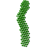
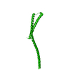
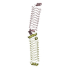
| タイトル | Crystal structure of a domain of a protein involved in formation of actin cytoskeleton |
|---|
 要素 要素 | Bud site selection protein 6 |
|---|
 キーワード キーワード | PROTEIN BINDING / coiled-coil |
|---|
| 機能・相同性 |  機能・相同性情報 機能・相同性情報
cytoskeletal regulatory protein binding / positive regulation of formin-nucleated actin cable assembly / polarisome / bipolar cellular bud site selection / budding cell apical bud growth / secretory vesicle / vesicle targeting / pseudohyphal growth / prospore membrane / establishment or maintenance of actin cytoskeleton polarity ...cytoskeletal regulatory protein binding / positive regulation of formin-nucleated actin cable assembly / polarisome / bipolar cellular bud site selection / budding cell apical bud growth / secretory vesicle / vesicle targeting / pseudohyphal growth / prospore membrane / establishment or maintenance of actin cytoskeleton polarity / positive regulation of actin nucleation / incipient cellular bud site / astral microtubule organization / cellular bud tip / cell tip / cellular bud neck / mating projection tip / cellular hyperosmotic response / spindle pole body / establishment of cell polarity / actin filament bundle assembly / regulation of actin cytoskeleton organization / enzyme activator activity / regulation of protein localization / actin binding / cytoplasm類似検索 - 分子機能 Actin interacting protein 3, C-terminal domain / Actin interacting protein 3, C-terminal / Actin interacting protein 3-like, C-terminal / : / : / Actin interacting protein 3 / Aip3p/Bud6 N-terminal domain / Actin interacting protein 3 / Methane Monooxygenase Hydroxylase; Chain G, domain 1 / Up-down Bundle / Mainly Alpha類似検索 - ドメイン・相同性 |
|---|
| 生物種 |   Saccharomyces cerevisiae (パン酵母) Saccharomyces cerevisiae (パン酵母) |
|---|
| 手法 |  X線回折 / シンクロトロン / 単波長異常分散 / 解像度: 2.904 Å X線回折 / シンクロトロン / 単波長異常分散 / 解像度: 2.904 Å |
|---|
 データ登録者 データ登録者 | Tu, D. / Eck, M.J. |
|---|
 引用 引用 | ジャーナル: Proc.Natl.Acad.Sci.USA / 年: 2012
タイトル: Structure of the formin-interaction domain of the actin nucleation-promoting factor Bud6.
著者: Tu, D. / Graziano, B.R. / Park, E. / Zheng, W. / Li, Y. / Goode, B.L. / Eck, M.J. |
|---|
| 履歴 | | 登録 | 2010年8月30日 | 登録サイト: RCSB / 処理サイト: RCSB |
|---|
| 改定 1.0 | 2011年10月12日 | Provider: repository / タイプ: Initial release |
|---|
| 改定 1.1 | 2012年12月5日 | Group: Database references |
|---|
| 改定 1.2 | 2012年12月26日 | Group: Database references |
|---|
| 改定 1.3 | 2024年11月6日 | Group: Data collection / Database references ...Data collection / Database references / Derived calculations / Structure summary
カテゴリ: chem_comp_atom / chem_comp_bond ...chem_comp_atom / chem_comp_bond / database_2 / pdbx_entry_details / pdbx_modification_feature / struct_conn / struct_ref_seq_dif
Item: _database_2.pdbx_DOI / _database_2.pdbx_database_accession ..._database_2.pdbx_DOI / _database_2.pdbx_database_accession / _struct_conn.pdbx_leaving_atom_flag / _struct_ref_seq_dif.details |
|---|
|
|---|
 ムービー
ムービー コントローラー


 データを開く
 基本情報
基本情報 構造の表示
構造の表示 ダウンロードとリンク
 その他のダウンロード


 PDBj
PDBj 集合体
集合体

 試料調製
試料調製 / ビームライン: 24-ID-C / 波長: 0.97934 Å
 解析
解析