 Movie
Movie Controller

[English] 日本語
 Yorodumi
- PDB-3one: Crystal structure of Lupinus luteus S-adenosyl-L-homocysteine hyd... -
+ Open data

- Basic information
| Entry | Database: PDB / ID: 3one | ||||||
|---|---|---|---|---|---|---|---|
| Title | Crystal structure of Lupinus luteus S-adenosyl-L-homocysteine hydrolase in complex with adenine | ||||||
 Components Components | Adenosylhomocysteinase | ||||||
 Keywords Keywords | HYDROLASE/HYDROLASE SUBSTRATE / Plant protein / enzyme-inhibitor complex / NAD cofactor / Regulation of SAM-dependent methylation reactions / HYDROLASE-HYDROLASE SUBSTRATE complex | ||||||
| Function / homology |  Function and homology information Function and homology informationadenosylhomocysteinase / adenosylhomocysteinase activity / L-methionine cycle / one-carbon metabolic process / cytosol Similarity search - Function | ||||||
| Biological species |  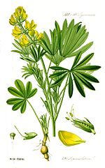 Lupinus luteus (yellow lupine) Lupinus luteus (yellow lupine) | ||||||
| Method |  X-RAY DIFFRACTION / SYNCHROTRON / MOLECULAR REPLACEMENT / Resolution: 1.35 Å X-RAY DIFFRACTION / SYNCHROTRON / MOLECULAR REPLACEMENT / Resolution: 1.35 Å | ||||||
 Authors Authors | Brzezinski, K. / Jaskolski, M. | ||||||
 Citation Citation | Journal: Acta Crystallogr.,Sect.D / Year: 2012 Title: High-resolution structures of complexes of plant S-adenosyl-L-homocysteine hydrolase (Lupinus luteus). Authors: Brzezinski, K. / Dauter, Z. / Jaskolski, M. #1: Journal: Acta Crystallogr.,Sect.F / Year: 2008 Title: Purification, crystallization and preliminary crystallographic studies of plant S-adenosyl-L-homocysteine hydrolase (Lupinus luteus). Authors: Brzezinski, K. / Bujacz, G. / Jaskolski, M. #2: Journal: Acta Biochim.Pol. / Year: 2001 Title: Sequence determination and analysis of S-adenosyl-L-homocysteine hydrolase from yellow lupine (Lupinus luteus). Authors: Brzezinski, K. / Janowski, R. / Podkowinski, J. / Jaskolski, M. #3: Journal: Nat.Struct.Biol. / Year: 1998Title: Structure determination of selenomethionyl S-adenosylhomocysteine hydrolase using data at a single wavelength. Authors: Turner, M.A. / Yuan, C.S. / Borchardt, R.T. / Hershfield, M.S. / Smith, G.D. / Howell, P.L. #4: Journal: Biochemistry / Year: 1999Title: Crystal structure of S-adenosylhomocysteine hydrolase from rat liver. Authors: Hu, Y. / Komoto, J. / Huang, Y. / Gomi, T. / Ogawa, H. / Takata, Y. / Fujioka, M. / Takusagawa, F. #5: Journal: J.Mol.Biol. / Year: 2004Title: Crystal structure of S-adenosyl-L-homocysteine hydrolase from the human malaria parasite Plasmodium falciparum. Authors: Tanaka, N. / Nakanishi, M. / Kusakabe, Y. / Shiraiwa, K. / Yabe, S. / Ito, Y. / Kitade, Y. / Nakamura, K.T. #6: Journal: Protein Sci. / Year: 2008Title: Crystal structures of Mycobacterium tuberculosis S-adenosyl-L-homocysteine hydrolase in ternary complex with substrate and inhibitors. Authors: Reddy, M.C. / Kuppan, G. / Shetty, N.D. / Owen, J.L. / Ioerger, T.R. / Sacchettini, J.C. #7: Journal: MOL.PHYLOGENET.EVOL. / Year: 2005 Title: Bayesian phylogenetic analysis reveals two-domain topology of S-adenosylhomocysteine hydrolase protein sequences. Authors: Stepkowski, T. / Brzezinski, K. / Legocki, A.B. / Jaskolski, M. / Bena, G. | ||||||
| History |
|
- Structure visualization
| Structure viewer | Molecule: MolmilJmol/JSmol |
|---|
- Downloads & links
-Download
| PDBx/mmCIF format | 3one.cif.gz | 448.3 KB | Display | PDBx/mmCIF format |
|---|---|---|---|---|
| PDB format | pdb3one.ent.gz | 366.3 KB | Display | PDB format |
| PDBx/mmJSON format | 3one.json.gz | Tree view | PDBx/mmJSON format | |
| Others |  Other downloads Other downloads |
-Validation report
| Arichive directory | https://data.pdbj.org/pub/pdb/validation_reports/on/3oneftp://data.pdbj.org/pub/pdb/validation_reports/on/3one | HTTPS FTP |
|---|
-Related structure data
| Related structure data | 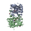 3ondC 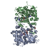 3onfC 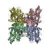 1v8bS S: Starting model for refinement C: citing same article ( |
|---|---|
| Similar structure data |


-Links
 PDBj
PDBj


- Assembly
| Deposited unit | 
| ||||||||
|---|---|---|---|---|---|---|---|---|---|
| 1 | 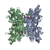
| ||||||||
| 2 |
| ||||||||
| Unit cell |
| ||||||||
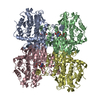
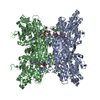
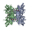
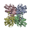
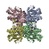
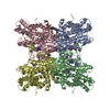
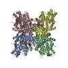
| Details | The asymmetric unit contains a dimer, which corresponds to the biological unit |
-Components
-Protein , 1 types, 2 molecules AB
| #1: Protein | Mass: 53673.336 Da / Num. of mol.: 2 Source method: isolated from a genetically manipulated source Source: (gene. exp.) Lupinus luteus (yellow lupine) / Gene: SAHH, SHH, shh-1 / Plasmid: pET15b / Production host:  |
|---|
-Non-polymers , 5 types, 1234 molecules 


| #2: Chemical |  Mass: 663.425 Da / Num. of mol.: 2 / Source method: obtained synthetically / Formula: C21H27N7O14P2 / Comment: NAD*YM Mass: 663.425 Da / Num. of mol.: 2 / Source method: obtained synthetically / Formula: C21H27N7O14P2 / Comment: NAD*YM#3: Chemical |  Mass: 122.143 Da / Num. of mol.: 3 / Source method: obtained synthetically / Formula: C4H12NO3 / Comment: pH buffer*YM Mass: 122.143 Da / Num. of mol.: 3 / Source method: obtained synthetically / Formula: C4H12NO3 / Comment: pH buffer*YM#4: Chemical |  Mass: 135.127 Da / Num. of mol.: 2 / Source method: obtained synthetically / Formula: C5H5N5 Mass: 135.127 Da / Num. of mol.: 2 / Source method: obtained synthetically / Formula: C5H5N5#5: Chemical |  Mass: 22.990 Da / Num. of mol.: 2 / Source method: obtained synthetically / Formula: Na Mass: 22.990 Da / Num. of mol.: 2 / Source method: obtained synthetically / Formula: Na#6: Water | ChemComp-HOH / | Mass: 18.015 Da / Num. of mol.: 1225 / Source method: isolated from a natural source / Formula: H2O |
|---|
-Experimental details
-Experiment
| Experiment | Method: X-RAY DIFFRACTION / Number of used crystals: 1 |
|---|
- Sample preparation
| Crystal | Density Matthews: 2.19 Å3/Da / Density % sol: 43.85 % |
|---|---|
| Crystal grow | Temperature: 292 K / Method: vapor diffusion, hanging drop / pH: 8 Details: 20% PEG 4000, 10% isopropanol, 0.1 M Tris-HCl pH 8.0, 2 mM 2'-deoxyadenosine, VAPOR DIFFUSION, HANGING DROP, temperature 292K |
-Data collection
| Diffraction | Mean temperature: 100 K |
|---|---|
| Diffraction source | Source: SYNCHROTRON / Site: EMBL/DESY, HAMBURG  / Beamline: X13 / Wavelength: 0.8086 / Beamline: X13 / Wavelength: 0.8086 |
| Detector | Type: MAR CCD 165 mm / Detector: CCD / Date: Dec 14, 2007 |
| Radiation | Monochromator: SI(111), HORIZONTALLY FOCUSING / Protocol: SINGLE WAVELENGTH / Monochromatic (M) / Laue (L): M / Scattering type: x-ray |
| Radiation wavelength | Wavelength: 0.8086 Å / Relative weight: 1 |
| Reflection | Resolution: 1.35→20 Å / Num. obs: 206827 / % possible obs: 99.3 % / Observed criterion σ(I): -3 / Redundancy: 9 % / Biso Wilson estimate: 14.4 Å2 / Rmerge(I) obs: 0.07 / Net I/σ(I): 28.1 |
| Reflection shell | Resolution: 1.35→1.37 Å / Redundancy: 6.2 % / Rmerge(I) obs: 0.67 / Mean I/σ(I) obs: 2.2 / % possible all: 98.2 |
- Processing
| Software |
| ||||||||||||||||||||||||||||||||||||||||||||||||||||||||||||||||||||||||||||||||||||||||||||||||||||||||||||||||||||||||||||||||||||||||||||||||||||||||||||||||||||||||||
|---|---|---|---|---|---|---|---|---|---|---|---|---|---|---|---|---|---|---|---|---|---|---|---|---|---|---|---|---|---|---|---|---|---|---|---|---|---|---|---|---|---|---|---|---|---|---|---|---|---|---|---|---|---|---|---|---|---|---|---|---|---|---|---|---|---|---|---|---|---|---|---|---|---|---|---|---|---|---|---|---|---|---|---|---|---|---|---|---|---|---|---|---|---|---|---|---|---|---|---|---|---|---|---|---|---|---|---|---|---|---|---|---|---|---|---|---|---|---|---|---|---|---|---|---|---|---|---|---|---|---|---|---|---|---|---|---|---|---|---|---|---|---|---|---|---|---|---|---|---|---|---|---|---|---|---|---|---|---|---|---|---|---|---|---|---|---|---|---|---|---|---|
| Refinement | Method to determine structure: MOLECULAR REPLACEMENT Starting model: 1V8B Resolution: 1.35→20 Å / Cor.coef. Fo:Fc: 0.982 / Cor.coef. Fo:Fc free: 0.973 / SU B: 1.582 / SU ML: 0.03 Isotropic thermal model: Anisotropic atomic displacement parameters Cross valid method: R FREE / ESU R: 0.048 / ESU R Free: 0.047 / Stereochemistry target values: MAXIMUM LIKELIHOOD Details: HYDROGEN ATOMS WERE ADDED AT RIDING POSITIONS. REFINEMENT OF INDIVIDUAL ANISOTROPIC ATOMIC DISPLACEMENT PARAMETERS (ADP)
| ||||||||||||||||||||||||||||||||||||||||||||||||||||||||||||||||||||||||||||||||||||||||||||||||||||||||||||||||||||||||||||||||||||||||||||||||||||||||||||||||||||||||||
| Solvent computation | Ion probe radii: 0.8 Å / Shrinkage radii: 0.8 Å / VDW probe radii: 1.4 Å / Solvent model: BABINET MODEL WITH MASK | ||||||||||||||||||||||||||||||||||||||||||||||||||||||||||||||||||||||||||||||||||||||||||||||||||||||||||||||||||||||||||||||||||||||||||||||||||||||||||||||||||||||||||
| Displacement parameters | Biso mean: 13.85 Å2
| ||||||||||||||||||||||||||||||||||||||||||||||||||||||||||||||||||||||||||||||||||||||||||||||||||||||||||||||||||||||||||||||||||||||||||||||||||||||||||||||||||||||||||
| Refinement step | Cycle: LAST / Resolution: 1.35→20 Å
| ||||||||||||||||||||||||||||||||||||||||||||||||||||||||||||||||||||||||||||||||||||||||||||||||||||||||||||||||||||||||||||||||||||||||||||||||||||||||||||||||||||||||||
| Refine LS restraints |
| ||||||||||||||||||||||||||||||||||||||||||||||||||||||||||||||||||||||||||||||||||||||||||||||||||||||||||||||||||||||||||||||||||||||||||||||||||||||||||||||||||||||||||
| LS refinement shell | Resolution: 1.35→1.39 Å / Total num. of bins used: 20
|
