 Movie
Movie Controller

[English] 日本語
 Yorodumi
- PDB-3ce6: Crystal structure of Mycobacterium tuberculosis S-adenosyl-L-homo... -
+ Open data

- Basic information
| Entry | Database: PDB / ID: 3ce6 | ||||||
|---|---|---|---|---|---|---|---|
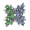
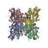
| Title | Crystal structure of Mycobacterium tuberculosis S-adenosyl-L-homocysteine hydrolase in ternary complex with NAD and adenosine | ||||||
 Components Components | Adenosylhomocysteinase | ||||||
 Keywords Keywords | HYDROLASE / Protein-substrate complex / dimer of dimers / NAD binding domain / 37 amino acid insertional region / One-carbon metabolism | ||||||
| Function / homology |  Function and homology information Function and homology informationadenosylhomocysteinase / L-methionine catabolic process / entry of bacterium into host cell / adhesion of symbiont to host cell / L-homocysteine biosynthetic process / adenosine metabolic process / adenosylhomocysteinase / adenosylhomocysteinase activity / L-methionine cycle / zymogen binding ...adenosylhomocysteinase / L-methionine catabolic process / entry of bacterium into host cell / adhesion of symbiont to host cell / L-homocysteine biosynthetic process / adenosine metabolic process / adenosylhomocysteinase / adenosylhomocysteinase activity / L-methionine cycle / zymogen binding / NAD+ binding / one-carbon metabolic process / peptidoglycan-based cell wall / extracellular region / plasma membrane / cytoplasm / cytosol Similarity search - Function | ||||||
| Biological species |   Mycobacterium tuberculosis (bacteria) Mycobacterium tuberculosis (bacteria) | ||||||
| Method |  X-RAY DIFFRACTION / SYNCHROTRON / MOLECULAR REPLACEMENT / Resolution: 1.6 Å X-RAY DIFFRACTION / SYNCHROTRON / MOLECULAR REPLACEMENT / Resolution: 1.6 Å | ||||||
 Authors Authors | Reddy, M.C.M. / Gokulan, K. / Shetty, N.D. / Owen, J.L. / Ioerger, T.R. / Sacchettini, J.C. | ||||||
 Citation Citation | Journal: Protein Sci. / Year: 2008 Title: Crystal structures of Mycobacterium tuberculosis S-adenosyl-L-homocysteine hydrolase in ternary complex with substrate and inhibitors. Authors: Reddy, M.C. / Kuppan, G. / Shetty, N.D. / Owen, J.L. / Ioerger, T.R. / Sacchettini, J.C. | ||||||
| History |
|
- Structure visualization
| Structure viewer | Molecule: MolmilJmol/JSmol |
|---|
- Downloads & links
-Download
| PDBx/mmCIF format | 3ce6.cif.gz | 422.8 KB | Display | PDBx/mmCIF format |
|---|---|---|---|---|
| PDB format | pdb3ce6.ent.gz | 340.8 KB | Display | PDB format |
| PDBx/mmJSON format | 3ce6.json.gz | Tree view | PDBx/mmJSON format | |
| Others |  Other downloads Other downloads |
-Validation report
| Arichive directory | https://data.pdbj.org/pub/pdb/validation_reports/ce/3ce6ftp://data.pdbj.org/pub/pdb/validation_reports/ce/3ce6 | HTTPS FTP |
|---|
-Related structure data
| Related structure data | 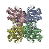 2zizC 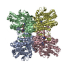 2zj0C  2zj1C  3dhyC 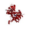 1li4S S: Starting model for refinement C: citing same article ( |
|---|---|
| Similar structure data |


-Links
 PDBj
PDBj
- Assembly
| Deposited unit | 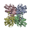
| ||||||||
|---|---|---|---|---|---|---|---|---|---|
| 1 |
| ||||||||
| Unit cell |
|
-Components
| #1: Protein | Mass: 54256.219 Da / Num. of mol.: 4 Source method: isolated from a genetically manipulated source Source: (gene. exp.) Mycobacterium tuberculosis (bacteria) / Strain: H37Rv / Gene: ahcY, sahH, Rv3248c, MT3346, MTCY20B11.23c / Plasmid: pET28TEV / Production host: References: UniProt: P60176, UniProt: P9WGV3*PLUS, adenosylhomocysteinase #2: Chemical | ChemComp-ADN /   Mass: 267.241 Da / Num. of mol.: 4 / Source method: obtained synthetically / Formula: C10H13N5O4 Mass: 267.241 Da / Num. of mol.: 4 / Source method: obtained synthetically / Formula: C10H13N5O4#3: Chemical | ChemComp-NAD /   Mass: 663.425 Da / Num. of mol.: 4 / Source method: obtained synthetically / Formula: C21H27N7O14P2 / Comment: NAD*YM Mass: 663.425 Da / Num. of mol.: 4 / Source method: obtained synthetically / Formula: C21H27N7O14P2 / Comment: NAD*YM#4: Water | ChemComp-HOH / |  Mass: 18.015 Da / Num. of mol.: 1736 / Source method: isolated from a natural source / Formula: H2O Mass: 18.015 Da / Num. of mol.: 1736 / Source method: isolated from a natural source / Formula: H2O |
|---|
-Experimental details
-Experiment
| Experiment | Method: X-RAY DIFFRACTION / Number of used crystals: 1 |
|---|
- Sample preparation
| Crystal | Density Matthews: 2.51 Å3/Da / Density % sol: 51.01 % |
|---|---|
| Crystal grow | Temperature: 291.5 K / pH: 8 Details: 20% PEG 1000, 200 mM Imidazole pH 8.0, 100 mM Calcium acetate, VAPOR DIFFUSION, HANGING DROP, temperature 291.5K |
-Data collection
| Diffraction | Mean temperature: 120 K |
|---|---|
| Diffraction source | Source: SYNCHROTRON / Site: APS  / Beamline: 19-ID / Wavelength: 0.964 / Beamline: 19-ID / Wavelength: 0.964 |
| Detector | Type: ADSC QUANTUM 315 / Detector: CCD / Date: Mar 4, 2005 / Details: MIRRORS |
| Radiation | Monochromator: SI(111) / Protocol: SINGLE WAVELENGTH / Monochromatic (M) / Laue (L): M / Scattering type: x-ray |
| Radiation wavelength | Wavelength: 0.964 Å / Relative weight: 1 |
| Reflection | Resolution: 1.5→100 Å / Num. obs: 308888 / % possible obs: 94.9 % / Observed criterion σ(I): 2 / Biso Wilson estimate: 17 Å2 / Rmerge(I) obs: 0.0487 / Rsym value: 0.0487 / Net I/σ(I): 13.1 |
| Reflection shell | Resolution: 1.5→1.54 Å / Rmerge(I) obs: 0.6821 / Mean I/σ(I) obs: 1.48 / Rsym value: 0.6821 / % possible all: 73.4 |
- Processing
| Software |
| ||||||||||||||||||||||||||||||||||||||||||||||||||||||||||||||||||||||||||||||||||||||||||||||||||||||||||||||||||||||||||||||||||||||||||||||||||||||||||||||||||||||||||
|---|---|---|---|---|---|---|---|---|---|---|---|---|---|---|---|---|---|---|---|---|---|---|---|---|---|---|---|---|---|---|---|---|---|---|---|---|---|---|---|---|---|---|---|---|---|---|---|---|---|---|---|---|---|---|---|---|---|---|---|---|---|---|---|---|---|---|---|---|---|---|---|---|---|---|---|---|---|---|---|---|---|---|---|---|---|---|---|---|---|---|---|---|---|---|---|---|---|---|---|---|---|---|---|---|---|---|---|---|---|---|---|---|---|---|---|---|---|---|---|---|---|---|---|---|---|---|---|---|---|---|---|---|---|---|---|---|---|---|---|---|---|---|---|---|---|---|---|---|---|---|---|---|---|---|---|---|---|---|---|---|---|---|---|---|---|---|---|---|---|---|---|
| Refinement | Method to determine structure: MOLECULAR REPLACEMENT Starting model: PDB ENTRY 1LI4 Resolution: 1.6→30 Å / Cor.coef. Fo:Fc: 0.95 / Cor.coef. Fo:Fc free: 0.935 / SU B: 1.799 / SU ML: 0.064 / Cross valid method: THROUGHOUT / ESU R: 0.097 / ESU R Free: 0.099 / Stereochemistry target values: MAXIMUM LIKELIHOOD / Details: HYDROGENS HAVE BEEN ADDED IN THE RIDING POSITIONS
| ||||||||||||||||||||||||||||||||||||||||||||||||||||||||||||||||||||||||||||||||||||||||||||||||||||||||||||||||||||||||||||||||||||||||||||||||||||||||||||||||||||||||||
| Solvent computation | Ion probe radii: 0.8 Å / Shrinkage radii: 0.8 Å / VDW probe radii: 1.4 Å / Solvent model: MASK | ||||||||||||||||||||||||||||||||||||||||||||||||||||||||||||||||||||||||||||||||||||||||||||||||||||||||||||||||||||||||||||||||||||||||||||||||||||||||||||||||||||||||||
| Displacement parameters | Biso mean: 18.37 Å2
| ||||||||||||||||||||||||||||||||||||||||||||||||||||||||||||||||||||||||||||||||||||||||||||||||||||||||||||||||||||||||||||||||||||||||||||||||||||||||||||||||||||||||||
| Refinement step | Cycle: LAST / Resolution: 1.6→30 Å
| ||||||||||||||||||||||||||||||||||||||||||||||||||||||||||||||||||||||||||||||||||||||||||||||||||||||||||||||||||||||||||||||||||||||||||||||||||||||||||||||||||||||||||
| Refine LS restraints |
| ||||||||||||||||||||||||||||||||||||||||||||||||||||||||||||||||||||||||||||||||||||||||||||||||||||||||||||||||||||||||||||||||||||||||||||||||||||||||||||||||||||||||||
| LS refinement shell | Resolution: 1.6→1.64 Å / Total num. of bins used: 20
|
