 Movie
Movie Controller

[English] 日本語
 Yorodumi
- PDB-3omw: Crystal structure of Ssu72, an essential eukaryotic phosphatase s... -
+ Open data

- Basic information
| Entry | Database: PDB / ID: 3omw | ||||||
|---|---|---|---|---|---|---|---|
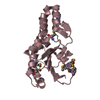
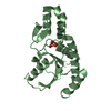
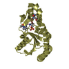
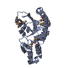
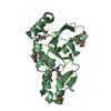
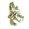
| Title | Crystal structure of Ssu72, an essential eukaryotic phosphatase specific for the C-terminal domain of RNA polymerase II | ||||||
 Components Components | CG14216 | ||||||
 Keywords Keywords | HYDROLASE / phosphatase / low molecular weight protein tyrosine phosphatase / C-terminal domain of RNA polymerase II / dephosphorylate C-terminal domain of RNA polymerase II / RNA polymerase II / transcription factor IIB / Pta1 / CPF complex | ||||||
| Function / homology |  Function and homology information Function and homology informationRNA polymerase II transcribes snRNA genes / RNA polymerase II CTD heptapeptide repeat phosphatase activity / mRNA cleavage and polyadenylation specificity factor complex / mRNA 3'-end processing / protein-serine/threonine phosphatase / phosphatase activity / termination of RNA polymerase II transcription / nucleus Similarity search - Function | ||||||
| Biological species |  | ||||||
| Method |  X-RAY DIFFRACTION / SYNCHROTRON / MOLECULAR REPLACEMENT / Resolution: 2.8701 Å X-RAY DIFFRACTION / SYNCHROTRON / MOLECULAR REPLACEMENT / Resolution: 2.8701 Å | ||||||
 Authors Authors | Zhang, Y. / Zhang, M. / Zhang, Y. | ||||||
 Citation Citation | Journal: Biochem.J. / Year: 2011 Title: Crystal structure of Ssu72, an essential eukaryotic phosphatase specific for the C-terminal domain of RNA polymerase II, in complex with a transition state analogue. Authors: Zhang, Y. / Zhang, M. / Zhang, Y. | ||||||
| History |
|
- Structure visualization
| Structure viewer | Molecule: MolmilJmol/JSmol |
|---|
- Downloads & links
-Download
| PDBx/mmCIF format | 3omw.cif.gz | 159 KB | Display | PDBx/mmCIF format |
|---|---|---|---|---|
| PDB format | pdb3omw.ent.gz | 127.7 KB | Display | PDB format |
| PDBx/mmJSON format | 3omw.json.gz | Tree view | PDBx/mmJSON format | |
| Others |  Other downloads Other downloads |
-Validation report
| Arichive directory | https://data.pdbj.org/pub/pdb/validation_reports/om/3omwftp://data.pdbj.org/pub/pdb/validation_reports/om/3omw | HTTPS FTP |
|---|
-Related structure data
| Related structure data |  3omxC  3fmvS S: Starting model for refinement C: citing same article ( |
|---|---|
| Similar structure data |


-Links
 PDBj
PDBj- Assembly
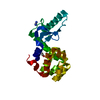
| Deposited unit | 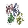
| ||||||||
|---|---|---|---|---|---|---|---|---|---|
| 1 | 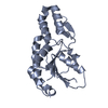
| ||||||||
| 2 | 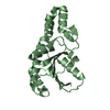
| ||||||||
| 3 | 
| ||||||||
| 4 | 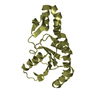
| ||||||||
| Unit cell |
|
-Components
| #1: Protein | Mass: 22232.393 Da / Num. of mol.: 4 Source method: isolated from a genetically manipulated source Source: (gene. exp.)  #2: Water | ChemComp-HOH / |  Mass: 18.015 Da / Num. of mol.: 15 / Source method: isolated from a natural source / Formula: H2O Mass: 18.015 Da / Num. of mol.: 15 / Source method: isolated from a natural source / Formula: H2O |
|---|
-Experimental details
-Experiment
| Experiment | Method: X-RAY DIFFRACTION / Number of used crystals: 1 |
|---|
- Sample preparation
| Crystal | Density Matthews: 2.97 Å3/Da / Density % sol: 58.64 % |
|---|---|
| Crystal grow | Temperature: 277 K / Method: vapor diffusion, sitting drop / pH: 7.5 Details: 0.1M HEPES pH 7.5, 10% isopropanol, 14-18% PEG4000, VAPOR DIFFUSION, SITTING DROP, temperature 277.0K |
-Data collection
| Diffraction | Mean temperature: 100 K |
|---|---|
| Diffraction source | Source: SYNCHROTRON / Site: ALS  / Beamline: 5.0.1 / Wavelength: 0.977 Å / Beamline: 5.0.1 / Wavelength: 0.977 Å |
| Detector | Type: ADSC QUANTUM 210 / Detector: CCD / Date: May 8, 2010 / Details: mirrors |
| Radiation | Monochromator: graphite / Protocol: SINGLE WAVELENGTH / Monochromatic (M) / Laue (L): M / Scattering type: x-ray |
| Radiation wavelength | Wavelength: 0.977 Å / Relative weight: 1 |
| Reflection | Resolution: 2.85→48.7 Å / Num. all: 26297 / Num. obs: 24877 / % possible obs: 94.6 % / Observed criterion σ(F): -3 / Observed criterion σ(I): 0 / Redundancy: 5 % / Rsym value: 0.087 / Net I/σ(I): 21.8 |
| Reflection shell | Resolution: 2.85→2.9 Å / Redundancy: 4.6 % / Mean I/σ(I) obs: 1.3 / Rsym value: 0.767 / % possible all: 96.2 |
- Processing
| Software |
| ||||||||||||||||||||||||||||||||||||||||||||||||||||||||||||||||||||||
|---|---|---|---|---|---|---|---|---|---|---|---|---|---|---|---|---|---|---|---|---|---|---|---|---|---|---|---|---|---|---|---|---|---|---|---|---|---|---|---|---|---|---|---|---|---|---|---|---|---|---|---|---|---|---|---|---|---|---|---|---|---|---|---|---|---|---|---|---|---|---|---|
| Refinement | Method to determine structure: MOLECULAR REPLACEMENT Starting model: PDB ENTRY 3FMV Resolution: 2.8701→48.507 Å / SU ML: 0.45 / σ(F): 1.34 / Phase error: 29.54 / Stereochemistry target values: ML
| ||||||||||||||||||||||||||||||||||||||||||||||||||||||||||||||||||||||
| Solvent computation | Shrinkage radii: 0.9 Å / VDW probe radii: 1.11 Å / Solvent model: FLAT BULK SOLVENT MODEL / Bsol: 53.682 Å2 / ksol: 0.312 e/Å3 | ||||||||||||||||||||||||||||||||||||||||||||||||||||||||||||||||||||||
| Displacement parameters |
| ||||||||||||||||||||||||||||||||||||||||||||||||||||||||||||||||||||||
| Refinement step | Cycle: LAST / Resolution: 2.8701→48.507 Å
| ||||||||||||||||||||||||||||||||||||||||||||||||||||||||||||||||||||||
| Refine LS restraints |
| ||||||||||||||||||||||||||||||||||||||||||||||||||||||||||||||||||||||
| LS refinement shell |
|
