 Movie
Movie Controller

[English] 日本語
 Yorodumi
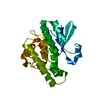
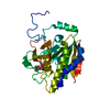
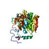
- PDB-3ois: Crystal Structure Xylellain, a cysteine protease from Xylella fas... -
+ Open data

- Basic information
| Entry | Database: PDB / ID: 3ois | ||||||
|---|---|---|---|---|---|---|---|
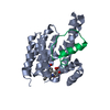
| Title | Crystal Structure Xylellain, a cysteine protease from Xylella fastidiosa | ||||||
 Components Components | Cysteine protease | ||||||
 Keywords Keywords | HYDROLASE / Alpha and beta | ||||||
| Function / homology |  Function and homology information Function and homology information | ||||||
| Biological species |  Xylella fastidiosa (bacteria) Xylella fastidiosa (bacteria) | ||||||
| Method |  X-RAY DIFFRACTION / SAD / Resolution: 1.65 Å X-RAY DIFFRACTION / SAD / Resolution: 1.65 Å | ||||||
 Authors Authors | Leite, N.R. / Faro, A.R. / Oliva, M.A.V. / Thiemann, O.H. / Oliva, G. | ||||||
 Citation Citation | Journal: Febs Lett. / Year: 2013 Title: The crystal structure of the cysteine protease Xylellain from Xylella fastidiosa reveals an intriguing activation mechanism. Authors: Leite, N.R. / Faro, A.R. / Dotta, M.A. / Faim, L.M. / Gianotti, A. / Silva, F.H. / Oliva, G. / Thiemann, O.H. | ||||||
| History |
|
- Structure visualization
| Structure viewer | Molecule: MolmilJmol/JSmol |
|---|
- Downloads & links
-Download
| PDBx/mmCIF format | 3ois.cif.gz | 266.9 KB | Display | PDBx/mmCIF format |
|---|---|---|---|---|
| PDB format | pdb3ois.ent.gz | 212.8 KB | Display | PDB format |
| PDBx/mmJSON format | 3ois.json.gz | Tree view | PDBx/mmJSON format | |
| Others |  Other downloads Other downloads |
-Validation report
| Arichive directory | https://data.pdbj.org/pub/pdb/validation_reports/oi/3oisftp://data.pdbj.org/pub/pdb/validation_reports/oi/3ois | HTTPS FTP |
|---|
-Related structure data
| Similar structure data |
|---|


-Links
 PDBj
PDBj


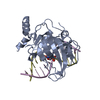
- Assembly
| Deposited unit | 
| ||||||||
|---|---|---|---|---|---|---|---|---|---|
| 1 | 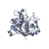
| ||||||||
| 2 | 
| ||||||||
| 3 | 
| ||||||||
| 4 | 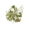
| ||||||||
| 5 | 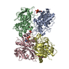
| ||||||||
| Unit cell |
|
-Components
| #1: Protein | Mass: 32977.129 Da / Num. of mol.: 4 Source method: isolated from a genetically manipulated source Source: (gene. exp.) Xylella fastidiosa (bacteria) / Gene: XF_0156 / Plasmid: pET28a / Production host: #2: Chemical | ChemComp-UDP /   Type: RNA linking / Mass: 404.161 Da / Num. of mol.: 4 / Source method: obtained synthetically / Formula: C9H14N2O12P2 / Comment: UDP*YM Type: RNA linking / Mass: 404.161 Da / Num. of mol.: 4 / Source method: obtained synthetically / Formula: C9H14N2O12P2 / Comment: UDP*YM#3: Water | ChemComp-HOH / |  Mass: 18.015 Da / Num. of mol.: 1435 / Source method: isolated from a natural source / Formula: H2O Mass: 18.015 Da / Num. of mol.: 1435 / Source method: isolated from a natural source / Formula: H2O |
|---|
-Experimental details
-Experiment
| Experiment | Method: X-RAY DIFFRACTION / Number of used crystals: 1 |
|---|
- Sample preparation
| Crystal | Density Matthews: 2.09 Å3/Da / Density % sol: 41.1 % |
|---|---|
| Crystal grow | Temperature: 298 K / Method: vapor diffusion, hanging drop / pH: 5.6 Details: 20-22% PEG 4000, 60 mM Sodium citrate, 134 mM Ammonium sulfate, pH 5.6, VAPOR DIFFUSION, HANGING DROP, temperature 298K |
-Data collection
| Diffraction | Mean temperature: 100 K | ||||||||||||||||||||||||||||||||||||||||||||||||||||||||||||||||||||||||||||||||||||||||
|---|---|---|---|---|---|---|---|---|---|---|---|---|---|---|---|---|---|---|---|---|---|---|---|---|---|---|---|---|---|---|---|---|---|---|---|---|---|---|---|---|---|---|---|---|---|---|---|---|---|---|---|---|---|---|---|---|---|---|---|---|---|---|---|---|---|---|---|---|---|---|---|---|---|---|---|---|---|---|---|---|---|---|---|---|---|---|---|---|---|
| Diffraction source | Source: ROTATING ANODE / Type: RIGAKU ULTRAX 18 / Wavelength: 1.5418 Å | ||||||||||||||||||||||||||||||||||||||||||||||||||||||||||||||||||||||||||||||||||||||||
| Detector | Type: MAR scanner 345 mm plate / Detector: IMAGE PLATE / Date: Mar 4, 2005 / Details: MIRROR | ||||||||||||||||||||||||||||||||||||||||||||||||||||||||||||||||||||||||||||||||||||||||
| Radiation | Protocol: SINGLE WAVELENGTH / Monochromatic (M) / Laue (L): M / Scattering type: x-ray | ||||||||||||||||||||||||||||||||||||||||||||||||||||||||||||||||||||||||||||||||||||||||
| Radiation wavelength | Wavelength: 1.5418 Å / Relative weight: 1 | ||||||||||||||||||||||||||||||||||||||||||||||||||||||||||||||||||||||||||||||||||||||||
| Reflection | Resolution: 1.65→78.664 Å / Num. all: 117070 / Num. obs: 117070 / % possible obs: 91 % / Observed criterion σ(F): 2 / Observed criterion σ(I): 2 / Redundancy: 8.1 % / Rmerge(I) obs: 0.054 / Rsym value: 0.054 / Net I/σ(I): 27 | ||||||||||||||||||||||||||||||||||||||||||||||||||||||||||||||||||||||||||||||||||||||||
| Reflection shell | Diffraction-ID: 1
|
-Phasing
| Phasing | Method: SAD |
|---|
- Processing
| Software |
| |||||||||||||||||||||||||||||||||||||||||||||||||||||||||||||||||
|---|---|---|---|---|---|---|---|---|---|---|---|---|---|---|---|---|---|---|---|---|---|---|---|---|---|---|---|---|---|---|---|---|---|---|---|---|---|---|---|---|---|---|---|---|---|---|---|---|---|---|---|---|---|---|---|---|---|---|---|---|---|---|---|---|---|---|
| Refinement | Method to determine structure: SAD / Resolution: 1.65→23.06 Å / Cor.coef. Fo:Fc: 0.971 / Cor.coef. Fo:Fc free: 0.949 / WRfactor Rfree: 0.2095 / WRfactor Rwork: 0.1595 / Occupancy max: 1 / Occupancy min: 0.01 / FOM work R set: 0.8597 / SU B: 2.286 / SU ML: 0.076 / SU R Cruickshank DPI: 0.1122 / SU Rfree: 0.1151 / Cross valid method: THROUGHOUT / σ(F): 0 / ESU R Free: 0.115 / Stereochemistry target values: MAXIMUM LIKELIHOOD Details: HYDROGENS HAVE BEEN ADDED IN THE RIDING POSITIONS U VALUES: REFINED INDIVIDUALLY
| |||||||||||||||||||||||||||||||||||||||||||||||||||||||||||||||||
| Solvent computation | Ion probe radii: 0.8 Å / Shrinkage radii: 0.8 Å / VDW probe radii: 1.4 Å / Solvent model: MASK | |||||||||||||||||||||||||||||||||||||||||||||||||||||||||||||||||
| Displacement parameters | Biso max: 93.77 Å2 / Biso mean: 22.8106 Å2 / Biso min: 4.92 Å2
| |||||||||||||||||||||||||||||||||||||||||||||||||||||||||||||||||
| Refinement step | Cycle: LAST / Resolution: 1.65→23.06 Å
| |||||||||||||||||||||||||||||||||||||||||||||||||||||||||||||||||
| Refine LS restraints |
| |||||||||||||||||||||||||||||||||||||||||||||||||||||||||||||||||
| LS refinement shell | Resolution: 1.65→1.693 Å / Total num. of bins used: 20
|
