 Movie
Movie Controller

[English] 日本語
 Yorodumi
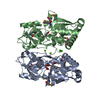
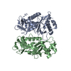
- PDB-3oiq: Crystal structure of yeast telomere protein Cdc13 OB1 and the cat... -
+ Open data

- Basic information
| Entry | Database: PDB / ID: 3oiq | ||||||
|---|---|---|---|---|---|---|---|
| Title | Crystal structure of yeast telomere protein Cdc13 OB1 and the catalytic subunit of DNA polymerase alpha Pol1 | ||||||
 Components Components |
| ||||||
 Keywords Keywords | PROTEIN BINDING / OB fold / dimer / dimeric complex | ||||||
| Function / homology |  Function and homology information Function and homology informationInhibition of replication initiation of damaged DNA by RB1/E2F1 / CST complex / H3-H4 histone complex chaperone activity / ribonucleoprotein complex localization / DNA replication initiation / Processive synthesis on the lagging strand / RNA-templated DNA biosynthetic process / Removal of the Flap Intermediate / translation elongation factor binding / Polymerase switching ...Inhibition of replication initiation of damaged DNA by RB1/E2F1 / CST complex / H3-H4 histone complex chaperone activity / ribonucleoprotein complex localization / DNA replication initiation / Processive synthesis on the lagging strand / RNA-templated DNA biosynthetic process / Removal of the Flap Intermediate / translation elongation factor binding / Polymerase switching / telomerase inhibitor activity / premeiotic DNA replication / regulation of telomere maintenance via telomerase / alpha DNA polymerase:primase complex / Activation of the pre-replicative complex / single-stranded telomeric DNA binding / lagging strand elongation / G-rich strand telomeric DNA binding / nuclear telomere cap complex / mitotic DNA replication initiation / telomere capping / leading strand elongation / DNA synthesis involved in DNA repair / DNA replication origin binding / DNA replication initiation / telomere maintenance via telomerase / telomere maintenance / replication fork / double-strand break repair / single-stranded DNA binding / 4 iron, 4 sulfur cluster binding / DNA-directed DNA polymerase / DNA-directed DNA polymerase activity / DNA replication / chromosome, telomeric region / cell division / nucleotide binding / chromatin binding / mitochondrion / zinc ion binding / identical protein binding Similarity search - Function | ||||||
| Biological species |  | ||||||
| Method |  X-RAY DIFFRACTION / SYNCHROTRON / MOLECULAR REPLACEMENT / Resolution: 2.4 Å X-RAY DIFFRACTION / SYNCHROTRON / MOLECULAR REPLACEMENT / Resolution: 2.4 Å | ||||||
 Authors Authors | Sun, J. / Yang, Y. / Wan, K. / Mao, N. / Yu, T.Y. / Lin, Y.C. / DeZwaan, D.C. / Freeman, B.C. / Lin, J.J. / Lue, N.F. / Lei, M. | ||||||
 Citation Citation | Journal: Cell Res. / Year: 2011 Title: Structural bases of dimerization of yeast telomere protein Cdc13 and its interaction with the catalytic subunit of DNA polymerase alpha. Authors: Sun, J. / Yang, Y. / Wan, K. / Mao, N. / Yu, T.Y. / Lin, Y.C. / DeZwaan, D.C. / Freeman, B.C. / Lin, J.J. / Lue, N.F. / Lei, M. | ||||||
| History |
|
- Structure visualization
| Structure viewer | Molecule: MolmilJmol/JSmol |
|---|
- Downloads & links
-Download
| PDBx/mmCIF format | 3oiq.cif.gz | 57.4 KB | Display | PDBx/mmCIF format |
|---|---|---|---|---|
| PDB format | pdb3oiq.ent.gz | 40.7 KB | Display | PDB format |
| PDBx/mmJSON format | 3oiq.json.gz | Tree view | PDBx/mmJSON format | |
| Others |  Other downloads Other downloads |
-Validation report
| Arichive directory | https://data.pdbj.org/pub/pdb/validation_reports/oi/3oiqftp://data.pdbj.org/pub/pdb/validation_reports/oi/3oiq | HTTPS FTP |
|---|
-Related structure data
| Related structure data |  3oipSC S: Starting model for refinement C: citing same article ( |
|---|---|
| Similar structure data |


-Links
 PDBj
PDBj


- Assembly
| Deposited unit | 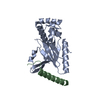
| ||||||||
|---|---|---|---|---|---|---|---|---|---|
| 1 | 
| ||||||||
| Unit cell |
|
-Components
| #1: Protein | Mass: 26596.436 Da / Num. of mol.: 1 Source method: isolated from a genetically manipulated source Source: (gene. exp.) Gene: CDC13, YDL220C / Production host:  |
|---|---|
| #2: Protein/peptide | Mass: 4116.635 Da / Num. of mol.: 1 Source method: isolated from a genetically manipulated source Source: (gene. exp.) Production host: |
| #3: Water | ChemComp-HOH /  Mass: 18.015 Da / Num. of mol.: 34 / Source method: isolated from a natural source / Formula: H2O Mass: 18.015 Da / Num. of mol.: 34 / Source method: isolated from a natural source / Formula: H2O |
-Experimental details
-Experiment
| Experiment | Method: X-RAY DIFFRACTION / Number of used crystals: 1 |
|---|
- Sample preparation
| Crystal | Density Matthews: 1.97 Å3/Da / Density % sol: 37.47 % |
|---|---|
| Crystal grow | Temperature: 277 K / Method: vapor diffusion, sitting drop / pH: 8 Details: 23% PEG3350, 0.2M Magnisium formate, 0.1M Tris-HCl, 5mM DTT, pH 8.0, VAPOR DIFFUSION, SITTING DROP, temperature 277K |
-Data collection
| Diffraction | Mean temperature: 100 K |
|---|---|
| Diffraction source | Source: SYNCHROTRON / Site: APS  / Beamline: 23-ID-D / Wavelength: 1.0782 / Beamline: 23-ID-D / Wavelength: 1.0782 |
| Detector | Type: MAR scanner 300 mm plate / Detector: IMAGE PLATE / Date: Apr 22, 2010 |
| Radiation | Monochromator: GRAPHITE / Protocol: SINGLE WAVELENGTH / Monochromatic (M) / Laue (L): M / Scattering type: x-ray |
| Radiation wavelength | Wavelength: 1.0782 Å / Relative weight: 1 |
| Reflection | Resolution: 2.4→100 Å / Num. all: 9997 / Num. obs: 9731 / % possible obs: 97.3 % / Observed criterion σ(F): 0 / Observed criterion σ(I): 0 |
- Processing
| Software |
| ||||||||||||||||||||
|---|---|---|---|---|---|---|---|---|---|---|---|---|---|---|---|---|---|---|---|---|---|
| Refinement | Method to determine structure: MOLECULAR REPLACEMENT Starting model: PDB ENTRY 3OIP Resolution: 2.4→50 Å / σ(F): 0 / Stereochemistry target values: Engh & Huber
| ||||||||||||||||||||
| Refinement step | Cycle: LAST / Resolution: 2.4→50 Å
| ||||||||||||||||||||
| Refine LS restraints |
|
