 Movie
Movie Controller


+ Open data

- Basic information
| Entry | Database: PDB / ID: 3ohp | ||||||
|---|---|---|---|---|---|---|---|
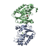
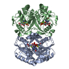
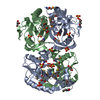
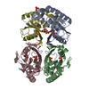
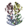
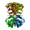
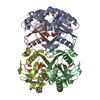
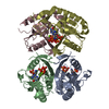
| Title | Crystal structure of HGPRT from Vibrio cholerae | ||||||
 Components Components | Hypoxanthine phosphoribosyltransferase | ||||||
 Keywords Keywords | TRANSFERASE / Structural Genomics / PSI-2 / Protein Structure Initiative / New York SGX Research Center for Structural Genomics / NYSGXRC / phosphoribosyltransferase | ||||||
| Function / homology |  Function and homology information Function and homology informationhypoxanthine phosphoribosyltransferase / guanine phosphoribosyltransferase activity / guanine salvage / hypoxanthine metabolic process / hypoxanthine phosphoribosyltransferase activity / GMP salvage / IMP salvage / purine ribonucleoside salvage / nucleotide binding / magnesium ion binding / cytosol Similarity search - Function | ||||||
| Biological species |   Vibrio cholerae (bacteria) Vibrio cholerae (bacteria) | ||||||
| Method |  X-RAY DIFFRACTION / SYNCHROTRON / MOLECULAR REPLACEMENT / Resolution: 2.04 Å X-RAY DIFFRACTION / SYNCHROTRON / MOLECULAR REPLACEMENT / Resolution: 2.04 Å | ||||||
 Authors Authors | Kim, J. / Ramagopal, U.A. / Almo, S.C. / Burley, S.K. / New York SGX Research Center for Structural Genomics (NYSGXRC) | ||||||
 Citation Citation | Journal: To be Published Title: Crystal structure of HGPRT from Vibrio cholerae Authors: Kim, J. / Ramagopal, U.A. / Almo, S.C. / Burley, S.K. | ||||||
| History |
|
- Structure visualization
| Structure viewer | Molecule: MolmilJmol/JSmol |
|---|
- Downloads & links
-Download
| PDBx/mmCIF format | 3ohp.cif.gz | 278.8 KB | Display | PDBx/mmCIF format |
|---|---|---|---|---|
| PDB format | pdb3ohp.ent.gz | 228.2 KB | Display | PDB format |
| PDBx/mmJSON format | 3ohp.json.gz | Tree view | PDBx/mmJSON format | |
| Others |  Other downloads Other downloads |
-Validation report
| Arichive directory | https://data.pdbj.org/pub/pdb/validation_reports/oh/3ohpftp://data.pdbj.org/pub/pdb/validation_reports/oh/3ohp | HTTPS FTP |
|---|
-Related structure data
| Related structure data | 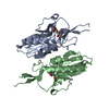 1g9sS S: Starting model for refinement |
|---|---|
| Similar structure data | |
| Other databases |

-Links
 PDBj
PDBj
- Assembly
| Deposited unit | 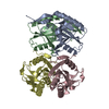
| ||||||||
|---|---|---|---|---|---|---|---|---|---|
| 1 |
| ||||||||
| Unit cell |
|
-Components
| #1: Protein | Mass: 20135.090 Da / Num. of mol.: 4 Source method: isolated from a genetically manipulated source Source: (gene. exp.) Vibrio cholerae (bacteria) / Gene: hpt, VC_0585 / Plasmid: LIC-PET30a / Production host: References: UniProt: Q9KUD7, hypoxanthine phosphoribosyltransferase #2: Water | ChemComp-HOH / |  Mass: 18.015 Da / Num. of mol.: 269 / Source method: isolated from a natural source / Formula: H2O Mass: 18.015 Da / Num. of mol.: 269 / Source method: isolated from a natural source / Formula: H2O |
|---|
-Experimental details
-Experiment
| Experiment | Method: X-RAY DIFFRACTION / Number of used crystals: 1 |
|---|
- Sample preparation
| Crystal | Density Matthews: 2.34 Å3/Da / Density % sol: 47.48 % |
|---|---|
| Crystal grow | Temperature: 293 K / Method: vapor diffusion, sitting drop / pH: 5.5 Details: 0.1M citrate PH 5.5, 20% PEG3000, VAPOR DIFFUSION, SITTING DROP, temperature 293K |
-Data collection
| Diffraction | Mean temperature: 100 K |
|---|---|
| Diffraction source | Source: SYNCHROTRON / Site: NSLS  / Beamline: X29A / Wavelength: 1.075 Å / Beamline: X29A / Wavelength: 1.075 Å |
| Detector | Type: ADSC QUANTUM 315 / Detector: CCD / Date: Feb 3, 2010 |
| Radiation | Monochromator: Double silicon(111) crystal / Protocol: SINGLE WAVELENGTH / Monochromatic (M) / Laue (L): M / Scattering type: x-ray |
| Radiation wavelength | Wavelength: 1.075 Å / Relative weight: 1 |
| Reflection | Resolution: 2.04→50 Å / Num. all: 47025 / Num. obs: 46597 / % possible obs: 99.1 % / Redundancy: 4 % / Biso Wilson estimate: 37.9 Å2 / Rmerge(I) obs: 0.07 / Rsym value: 0.058 / Net I/σ(I): 17.8 |
| Reflection shell | Resolution: 2.04→2.11 Å / Redundancy: 3.7 % / Rmerge(I) obs: 0.353 / Mean I/σ(I) obs: 3.4 / Num. unique all: 4386 / Rsym value: 0.294 / % possible all: 92.6 |
- Processing
| Software |
| |||||||||||||||||||||||||||||||||||||||||||||||||||||||||||||||||||||||||||||||||||||||||||||||||||||||||||||||||||||||||||||
|---|---|---|---|---|---|---|---|---|---|---|---|---|---|---|---|---|---|---|---|---|---|---|---|---|---|---|---|---|---|---|---|---|---|---|---|---|---|---|---|---|---|---|---|---|---|---|---|---|---|---|---|---|---|---|---|---|---|---|---|---|---|---|---|---|---|---|---|---|---|---|---|---|---|---|---|---|---|---|---|---|---|---|---|---|---|---|---|---|---|---|---|---|---|---|---|---|---|---|---|---|---|---|---|---|---|---|---|---|---|---|---|---|---|---|---|---|---|---|---|---|---|---|---|---|---|---|
| Refinement | Method to determine structure: MOLECULAR REPLACEMENT Starting model: 1G9S Resolution: 2.04→50 Å / Cor.coef. Fo:Fc: 0.961 / Cor.coef. Fo:Fc free: 0.939 / SU B: 9.576 / SU ML: 0.118 / Cross valid method: THROUGHOUT / ESU R: 0.191 / ESU R Free: 0.167 / Stereochemistry target values: MAXIMUM LIKELIHOOD / Details: HYDROGENS HAVE BEEN ADDED IN THE RIDING POSITIONS
| |||||||||||||||||||||||||||||||||||||||||||||||||||||||||||||||||||||||||||||||||||||||||||||||||||||||||||||||||||||||||||||
| Solvent computation | Ion probe radii: 0.8 Å / Shrinkage radii: 0.8 Å / VDW probe radii: 1.4 Å / Solvent model: MASK | |||||||||||||||||||||||||||||||||||||||||||||||||||||||||||||||||||||||||||||||||||||||||||||||||||||||||||||||||||||||||||||
| Displacement parameters | Biso mean: 32.106 Å2
| |||||||||||||||||||||||||||||||||||||||||||||||||||||||||||||||||||||||||||||||||||||||||||||||||||||||||||||||||||||||||||||
| Refinement step | Cycle: LAST / Resolution: 2.04→50 Å
| |||||||||||||||||||||||||||||||||||||||||||||||||||||||||||||||||||||||||||||||||||||||||||||||||||||||||||||||||||||||||||||
| Refine LS restraints |
| |||||||||||||||||||||||||||||||||||||||||||||||||||||||||||||||||||||||||||||||||||||||||||||||||||||||||||||||||||||||||||||
| LS refinement shell | Resolution: 2.038→2.091 Å / Total num. of bins used: 20
| |||||||||||||||||||||||||||||||||||||||||||||||||||||||||||||||||||||||||||||||||||||||||||||||||||||||||||||||||||||||||||||
| Refinement TLS params. | Method: refined / Refine-ID: X-RAY DIFFRACTION
| |||||||||||||||||||||||||||||||||||||||||||||||||||||||||||||||||||||||||||||||||||||||||||||||||||||||||||||||||||||||||||||
| Refinement TLS group |
|
