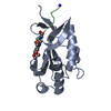
- PDB-3obx: Crystal structure of the Tsg101 UEV domain in complex with a HIV-... -
+
Open data
ID or keywords:
Loading...
-
Basic information
Entry
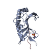
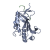
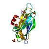
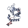
Database: PDB / ID: 3obx
Title
Crystal structure of the Tsg101 UEV domain in complex with a HIV-1 Gag P7A mutant peptide
Components
Gag polyprotein
Tumor susceptibility gene 101 protein
Keywords
PROTEIN TRANSPORT / Protein tranport / ubiquitin / HIV-1 Gag
Function / homology
Function and homology information
positive regulation of viral budding via host ESCRT complex / positive regulation of ubiquitin-dependent endocytosis / extracellular transport / ESCRT I complex / negative regulation of epidermal growth factor-activated receptor activity / regulation of extracellular exosome assembly / viral budding / regulation of MAP kinase activity / exosomal secretion / protein transport to vacuole involved in ubiquitin-dependent protein catabolic process via the multivesicular body sorting pathway ...positive regulation of viral budding via host ESCRT complex / positive regulation of ubiquitin-dependent endocytosis / extracellular transport / ESCRT I complex / negative regulation of epidermal growth factor-activated receptor activity / regulation of extracellular exosome assembly / viral budding / regulation of MAP kinase activity / exosomal secretion / protein transport to vacuole involved in ubiquitin-dependent protein catabolic process via the multivesicular body sorting pathway / membrane fission / positive regulation of exosomal secretion / multivesicular body assembly / ubiquitin-dependent protein catabolic process via the multivesicular body sorting pathway / Flemming body / virion binding / endosome to lysosome transport / viral budding via host ESCRT complex / negative regulation of epidermal growth factor receptor signaling pathway / viral release from host cell / autophagosome maturation / keratinocyte differentiation / multivesicular body / Endosomal Sorting Complex Required For Transport (ESCRT) / Membrane binding and targetting of GAG proteins / HCMV Late Events / macroautophagy / ubiquitin binding / regulation of cell growth / Late endosomal microautophagy / Budding and maturation of HIV virion / host multivesicular body / calcium-dependent protein binding / transcription corepressor activity / late endosome / late endosome membrane / viral nucleocapsid / early endosome membrane / early endosome / endosome / regulation of cell cycle / endosome membrane / viral translational frameshifting / negative regulation of cell population proliferation / cell division / ubiquitin protein ligase binding / centrosome / host cell nucleus / host cell plasma membrane / protein-containing complex binding / nucleolus / virion membrane / structural molecule activity / negative regulation of transcription by RNA polymerase II / protein homodimerization activity / DNA binding / RNA binding / extracellular exosome / zinc ion binding / plasma membrane / cytoplasm / cytosol Similarity search - Function
In the structure databanks used in Yorodumi, some data are registered as the other names, "COVID-19 virus" and "2019-nCoV". Here are the details of the virus and the list of structure data.
Jan 31, 2019. EMDB accession codes are about to change! (news from PDBe EMDB page)
EMDB accession codes are about to change! (news from PDBe EMDB page)
The allocation of 4 digits for EMDB accession codes will soon come to an end. Whilst these codes will remain in use, new EMDB accession codes will include an additional digit and will expand incrementally as the available range of codes is exhausted. The current 4-digit format prefixed with “EMD-” (i.e. EMD-XXXX) will advance to a 5-digit format (i.e. EMD-XXXXX), and so on. It is currently estimated that the 4-digit codes will be depleted around Spring 2019, at which point the 5-digit format will come into force.
The EM Navigator/Yorodumi systems omit the EMD- prefix.
Related info.:Q: What is EMD? / ID/Accession-code notation in Yorodumi/EM Navigator
Yorodumi is a browser for structure data from EMDB, PDB, SASBDB, etc.
This page is also the successor to EM Navigator detail page, and also detail information page/front-end page for Omokage search.
The word "yorodu" (or yorozu) is an old Japanese word meaning "ten thousand". "mi" (miru) is to see.
Related info.:EMDB / PDB / SASBDB / Comparison of 3 databanks / Yorodumi Search / Aug 31, 2016. New EM Navigator & Yorodumi / Yorodumi Papers / Jmol/JSmol / Function and homology information / Changes in new EM Navigator and Yorodumi
 Movie
Movie Controller

 Yorodumi
 Open data
 Basic information
 Components
 Keywords
 Function and homology information
 Homo sapiens (human)
 X-RAY DIFFRACTION /
  Authors
 Citation
 Structure visualization
 Downloads & links
 Other downloads


 PDBj
PDBj


 Assembly

 Mass: 18.015 Da / Num. of mol.: 123 / Source method: isolated from a natural source / Formula: H2O
 Sample preparation
 / Beamline: 22-ID / Wavelength: 1 Å
 Processing