 Movie
Movie Controller

[English] 日本語
 Yorodumi
- PDB-3ob0: A non-self sugar mimic of the HIV glycan shield shows enhanced an... -
+ Open data

- Basic information
| Entry | Database: PDB / ID: 3ob0 | |||||||||
|---|---|---|---|---|---|---|---|---|---|---|
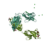
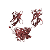
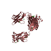
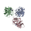
| Title | A non-self sugar mimic of the HIV glycan shield shows enhanced antigenicity | |||||||||
 Components Components |
| |||||||||
 Keywords Keywords | IMMUNE SYSTEM / Fab | |||||||||
| Function / homology | Immunoglobulins / Immunoglobulin-like / Sandwich / Mainly Beta Function and homology information Function and homology information | |||||||||
| Biological species |  Homo sapiens (human) Homo sapiens (human) | |||||||||
| Method |  X-RAY DIFFRACTION / SYNCHROTRON / MOLECULAR REPLACEMENT / Resolution: 2.85 Å X-RAY DIFFRACTION / SYNCHROTRON / MOLECULAR REPLACEMENT / Resolution: 2.85 Å | |||||||||
 Authors Authors | Doores, K.J. / Fulton, Z. / Hong, V. / Patel, M.K. / Scanlan, C.N. / Wormald, M.R. / Finn, M.G. / Burton, D.R. / Wilson, I.A. / Davis, B.G. | |||||||||
 Citation Citation | Journal: Proc.Natl.Acad.Sci.USA / Year: 2010 Title: A nonself sugar mimic of the HIV glycan shield shows enhanced antigenicity. Authors: Doores, K.J. / Fulton, Z. / Hong, V. / Patel, M.K. / Scanlan, C.N. / Wormald, M.R. / Finn, M.G. / Burton, D.R. / Wilson, I.A. / Davis, B.G. | |||||||||
| History |
|
- Structure visualization
| Structure viewer | Molecule: MolmilJmol/JSmol |
|---|
- Downloads & links
-Download
| PDBx/mmCIF format | 3ob0.cif.gz | 175.5 KB | Display | PDBx/mmCIF format |
|---|---|---|---|---|
| PDB format | pdb3ob0.ent.gz | 138.7 KB | Display | PDB format |
| PDBx/mmJSON format | 3ob0.json.gz | Tree view | PDBx/mmJSON format | |
| Others |  Other downloads Other downloads |
-Validation report
| Arichive directory | https://data.pdbj.org/pub/pdb/validation_reports/ob/3ob0ftp://data.pdbj.org/pub/pdb/validation_reports/ob/3ob0 | HTTPS FTP |
|---|
-Related structure data
| Related structure data |  3oayC  3oazC  1op3 C: citing same article ( S: Starting model for refinement |
|---|---|
| Similar structure data |


-Links
 PDBj
PDBj

- Assembly
| Deposited unit | 
| ||||||||
|---|---|---|---|---|---|---|---|---|---|
| 1 | 
| ||||||||
| 2 | 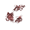
| ||||||||
| Unit cell |
|
-Components
| #1: Antibody | Mass: 23201.840 Da / Num. of mol.: 2 / Fragment: Fab Source method: isolated from a genetically manipulated source Source: (gene. exp.) Homo sapiens (human) / Organ (production host): ovary / Production host:   Cricetulus griseus (Chinese hamster) Cricetulus griseus (Chinese hamster)#2: Antibody | Mass: 23845.791 Da / Num. of mol.: 2 Source method: isolated from a genetically manipulated source Source: (gene. exp.) Homo sapiens (human) / Organ (production host): ovary / Production host: Cricetulus griseus (Chinese hamster)#3: Polysaccharide | Source method: isolated from a genetically manipulated source #4: Water | ChemComp-HOH / |  Mass: 18.015 Da / Num. of mol.: 8 / Source method: isolated from a natural source / Formula: H2O Mass: 18.015 Da / Num. of mol.: 8 / Source method: isolated from a natural source / Formula: H2OHas protein modification | Y | |
|---|
-Experimental details
-Experiment
| Experiment | Method: X-RAY DIFFRACTION / Number of used crystals: 1 |
|---|
- Sample preparation
| Crystal | Density Matthews: 2.64 Å3/Da / Density % sol: 53.46 % |
|---|
-Data collection
| Diffraction source | Source: SYNCHROTRON / Site: ALS  / Beamline: 8.2.1 / Beamline: 8.2.1 |
|---|---|
| Radiation | Protocol: SINGLE WAVELENGTH / Monochromatic (M) / Laue (L): M / Scattering type: x-ray |
| Radiation wavelength | Relative weight: 1 |
| Reflection | Resolution: 2.85→30 Å / Num. obs: 21555 / Biso Wilson estimate: 59.67 Å2 |
- Processing
| Software |
| ||||||||||||||||||||
|---|---|---|---|---|---|---|---|---|---|---|---|---|---|---|---|---|---|---|---|---|---|
| Refinement | Method to determine structure: MOLECULAR REPLACEMENT Starting model: PDB entry 1OP3 1op3 Resolution: 2.85→30 Å / Cor.coef. Fo:Fc: 0.8658 / Cor.coef. Fo:Fc free: 0.8403 / Occupancy max: 1 / Occupancy min: 1 / Cross valid method: THROUGHOUT / σ(F): 0
| ||||||||||||||||||||
| Displacement parameters | Biso max: 229.74 Å2 / Biso mean: 79.1252 Å2 / Biso min: 36.04 Å2
| ||||||||||||||||||||
| Refine analyze | Luzzati coordinate error obs: 0.496 Å | ||||||||||||||||||||
| Refinement step | Cycle: LAST / Resolution: 2.85→30 Å
| ||||||||||||||||||||
| LS refinement shell | Resolution: 2.85→2.99 Å / Total num. of bins used: 11
|
