Type: MARMOSAIC 225 mm CCD / Detector: CCD / Date: May 11, 2010 / Details: Double crystal monochromator
Radiation
Monochromator: double crystal monochromator / Protocol: SINGLE WAVELENGTH / Monochromatic (M) / Laue (L): M / Scattering type: x-ray
Radiation wavelength
Wavelength: 0.97904 Å / Relative weight: 1
Reflection twin
Crystal-ID
ID
Operator
Domain-ID
Fraction
1
1
H, K, L
1
0.941
1
1
K, H, -L
2
0.059
Reflection
Resolution: 2.5→50 Å / Num. obs: 6291 / % possible obs: 99.9 % / Redundancy: 3.9 % / Rmerge(I) obs: 0.074 / Χ2: 3.016 / Net I/σ(I): 19.1
Reflection shell
Resolution (Å)
Redundancy (%)
Rmerge(I) obs
Num. unique all
Χ2
% possible all
2.5-2.59
3.9
0.549
641
1.031
100
2.59-2.69
4
0.426
626
1.011
100
2.69-2.82
3.9
0.322
613
1.117
100
2.82-2.96
3.9
0.236
634
1.323
100
2.96-3.15
4
0.148
630
1.508
100
3.15-3.39
3.9
0.087
627
1.93
100
3.39-3.73
4
0.066
638
1.902
100
3.73-4.27
4
0.064
629
1.636
100
4.27-5.38
4
0.056
625
4.988
100
5.38-50
4
0.067
628
13.674
99.2
-
Phasing
Phasing
Method: SAD
Phasing MAD
D res high: 2.5 Å / D res low: 35.93 Å / FOM acentric: 0.346 / FOM centric: 0 / Reflection acentric: 6265 / Reflection centric: 0
Phasing MAD set
ID
R cullis acentric
R cullis centric
Highest resolution (Å)
Lowest resolution (Å)
Reflection acentric
Reflection centric
ISO_1
0
0
2.5
35.93
6265
0
ANO_1
0.595
0
2.5
35.93
6261
0
Phasing MAD set shell
ID
Resolution (Å)
R cullis acentric
R cullis centric
Reflection acentric
Reflection centric
ISO_1
9.66-35.93
0
0
105
0
ISO_1
6.95-9.66
0
0
186
0
ISO_1
5.71-6.95
0
0
223
0
ISO_1
4.96-5.71
0
0
287
0
ISO_1
4.45-4.96
0
0
309
0
ISO_1
4.07-4.45
0
0
344
0
ISO_1
3.77-4.07
0
0
371
0
ISO_1
3.53-3.77
0
0
410
0
ISO_1
3.33-3.53
0
0
426
0
ISO_1
3.16-3.33
0
0
453
0
ISO_1
3.01-3.16
0
0
457
0
ISO_1
2.88-3.01
0
0
493
0
ISO_1
2.77-2.88
0
0
526
0
ISO_1
2.67-2.77
0
0
551
0
ISO_1
2.58-2.67
0
0
541
0
ISO_1
2.5-2.58
0
0
583
0
ANO_1
9.66-35.93
0.287
0
103
0
ANO_1
6.95-9.66
0.229
0
186
0
ANO_1
5.71-6.95
0.274
0
223
0
ANO_1
4.96-5.71
0.366
0
287
0
ANO_1
4.45-4.96
0.402
0
309
0
ANO_1
4.07-4.45
0.581
0
344
0
ANO_1
3.77-4.07
0.671
0
370
0
ANO_1
3.53-3.77
0.649
0
410
0
ANO_1
3.33-3.53
0.734
0
426
0
ANO_1
3.16-3.33
0.787
0
453
0
ANO_1
3.01-3.16
0.875
0
457
0
ANO_1
2.88-3.01
0.929
0
493
0
ANO_1
2.77-2.88
0.949
0
526
0
ANO_1
2.67-2.77
0.978
0
551
0
ANO_1
2.58-2.67
0.994
0
541
0
ANO_1
2.5-2.58
1.002
0
582
0
Phasing MAD set site
ID
Cartn x (Å)
Cartn y (Å)
Cartn z (Å)
Atom type symbol
B iso
Occupancy
1
-24.199
58.232
29.627
SE
81.05
0.91
2
-24.816
58.459
39.46
SE
72.42
0.88
Phasing MAD shell
Resolution (Å)
FOM acentric
FOM centric
Reflection acentric
Reflection centric
9.66-35.93
0.611
0
105
0
6.95-9.66
0.649
0
186
0
5.71-6.95
0.617
0
223
0
4.96-5.71
0.556
0
287
0
4.45-4.96
0.553
0
309
0
4.07-4.45
0.473
0
344
0
3.77-4.07
0.4
0
371
0
3.53-3.77
0.45
0
410
0
3.33-3.53
0.411
0
426
0
3.16-3.33
0.378
0
453
0
3.01-3.16
0.307
0
457
0
2.88-3.01
0.274
0
493
0
2.77-2.88
0.238
0
526
0
2.67-2.77
0.189
0
551
0
2.58-2.67
0.157
0
541
0
2.5-2.58
0.137
0
583
0
Phasing dm
Method: Solvent flattening and Histogram matching / Reflection: 6263
Phasing dm shell
Resolution (Å)
Delta phi final
FOM
Reflection
5.76-100
55.6
0.847
503
4.6-5.76
51
0.934
511
4.01-4.6
57.8
0.937
502
3.65-4.01
57.5
0.909
502
3.39-3.65
58.6
0.898
502
3.19-3.39
67.4
0.889
511
3.03-3.19
72.8
0.886
511
2.89-3.03
75.9
0.871
507
2.78-2.89
81
0.794
505
2.68-2.78
76.7
0.826
503
2.6-2.68
82.2
0.809
509
2.5-2.6
85.8
0.738
697
-
Processing
Software
Name
Version
Classification
NB
DENZO
datareduction
SCALEPACK
datascaling
SHARP
phasing
DM
6.1
phasing
REFMAC
refmac_5.6.0081
refinement
PDB_EXTRACT
3.1
dataextraction
MxDC
datacollection
HKL-2000
datareduction
HKL-2000
datascaling
Refinement
Method to determine structure: SAD / Resolution: 2.5→35.93 Å / Cor.coef. Fo:Fc: 0.947 / Cor.coef. Fo:Fc free: 0.938 / WRfactor Rfree: 0.265 / WRfactor Rwork: 0.232 / Occupancy max: 1 / Occupancy min: 1 / SU B: 10.86 / SU ML: 0.205 / Cross valid method: THROUGHOUT / σ(F): 0 / ESU R: 0.063 / ESU R Free: 0.052 / Stereochemistry target values: MAXIMUM LIKELIHOOD Details: HYDROGENS HAVE BEEN USED IF PRESENT IN THE INPUT U VALUES : REFINED INDIVIDUALLY, The data is hemohedral twinning with twinning operators: ((H, K, L),(K, H, -L) and corresponding twinned fractions: 0.941, 0.059.
Rfactor
Num. reflection
% reflection
Selection details
Rfree
0.2612
288
4.6 %
RANDOM
Rwork
0.2183
-
-
-
obs
0.2201
6288
99.89 %
-
Solvent computation
Ion probe radii: 0.8 Å / Shrinkage radii: 0.8 Å / VDW probe radii: 1.2 Å / Solvent model: BABINET MODEL WITH MASK
In the structure databanks used in Yorodumi, some data are registered as the other names, "COVID-19 virus" and "2019-nCoV". Here are the details of the virus and the list of structure data.
Jan 31, 2019. EMDB accession codes are about to change! (news from PDBe EMDB page)
EMDB accession codes are about to change! (news from PDBe EMDB page)
The allocation of 4 digits for EMDB accession codes will soon come to an end. Whilst these codes will remain in use, new EMDB accession codes will include an additional digit and will expand incrementally as the available range of codes is exhausted. The current 4-digit format prefixed with “EMD-” (i.e. EMD-XXXX) will advance to a 5-digit format (i.e. EMD-XXXXX), and so on. It is currently estimated that the 4-digit codes will be depleted around Spring 2019, at which point the 5-digit format will come into force.
The EM Navigator/Yorodumi systems omit the EMD- prefix.
Related info.:Q: What is EMD? / ID/Accession-code notation in Yorodumi/EM Navigator
Yorodumi is a browser for structure data from EMDB, PDB, SASBDB, etc.
This page is also the successor to EM Navigator detail page, and also detail information page/front-end page for Omokage search.
The word "yorodu" (or yorozu) is an old Japanese word meaning "ten thousand". "mi" (miru) is to see.
Related info.:EMDB / PDB / SASBDB / Comparison of 3 databanks / Yorodumi Search / Aug 31, 2016. New EM Navigator & Yorodumi / Yorodumi Papers / Jmol/JSmol / Function and homology information / Changes in new EM Navigator and Yorodumi
 Movie
Movie Controller


 Open data
 Basic information
 Components
 Keywords
 Function and homology information
 Homo sapiens (human)
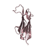
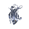
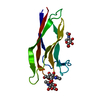
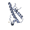
 X-RAY DIFFRACTION /
  Authors
 Citation
 Structure visualization
 Downloads & links
 Other downloads


 PDBj
PDBj
 Assembly

 Mass: 18.015 Da / Num. of mol.: 1 / Source method: isolated from a natural source / Formula: H2O
 Sample preparation
 / Beamline: 08ID-1 / Wavelength: 0.97904 Å
 Processing