Mass: 18.015 Da / Num. of mol.: 107 / Source method: isolated from a natural source / Formula: H2O
Sequence details
THE FULL SEQUENCE OF THIS PROTEIN USED FOR CRYSTALLIZATION IS ...THE FULL SEQUENCE OF THIS PROTEIN USED FOR CRYSTALLIZATION IS MGSSHHHHHHSSGVRGSHMKLSDIARLAGVSVTTASYVINGKAEQQRISNST VERVRAVVEAHGFTPNPQAAGLRSRHTRTLGFILPDLENPSYARIAKQLEQGARARGYQL LIASSDDQPDSERQLQQLFRARRCDALFVASCLPPEDDSYRELQDKGLPVIAIDRRLDPA HFCSVISDDRDASRQLAASLLSSAPRSIALIGARPELSVSQARAGGFDEALQGYTGEVRR YQGEAFSRECGQRLMQQLIDDLGGLPDALVTTSYVLLQGVFDTLQARPVDSRQLQL GTFGDNQLLDFLPLPVNAMAQQHGQIAATALELALAAIEEKRYEPGVHAVGRTFKQ RISVA. HOWEVER, RESIDUES AT N-TERMINUS SEEM TO BE DEGRADED IN CRYSTAL AND THE ACTUAL CRYSTALLIZED PROTEIN SEQUENCE MAY BE SHORTER. IN FACT MASS SPECTROMETRY COULD ONLY SHOW A PEAK OF 30.4KDA WEIGHT. ONLY THE ORDERED PART OF THE PROTEIN BY THE ELECTRON DENSITY MAP IS REPORTED IN SEQRES RECORDS. THE FIRST 18 RESIDUES MGSSHHHHHHSSGVRGSH ARE EXPRESSION TAG SEQUENCE.
-
Experimental details
-
Experiment
Experiment
Method: X-RAY DIFFRACTION / Number of used crystals: 1
-
Sample preparation
Crystal
Density Matthews: 2.23 Å3/Da / Density % sol: 44.93 %
Crystal grow
Temperature: 294 K / Method: vapor diffusion / pH: 6.5 Details: 0.1M Mes pH 6.5, 0.2M NaAc, 15 % PEG 8000, VAPOR DIFFUSION, temperature 294K
In the structure databanks used in Yorodumi, some data are registered as the other names, "COVID-19 virus" and "2019-nCoV". Here are the details of the virus and the list of structure data.
Jan 31, 2019. EMDB accession codes are about to change! (news from PDBe EMDB page)
EMDB accession codes are about to change! (news from PDBe EMDB page)
The allocation of 4 digits for EMDB accession codes will soon come to an end. Whilst these codes will remain in use, new EMDB accession codes will include an additional digit and will expand incrementally as the available range of codes is exhausted. The current 4-digit format prefixed with “EMD-” (i.e. EMD-XXXX) will advance to a 5-digit format (i.e. EMD-XXXXX), and so on. It is currently estimated that the 4-digit codes will be depleted around Spring 2019, at which point the 5-digit format will come into force.
The EM Navigator/Yorodumi systems omit the EMD- prefix.
Related info.:Q: What is EMD? / ID/Accession-code notation in Yorodumi/EM Navigator
Yorodumi is a browser for structure data from EMDB, PDB, SASBDB, etc.
This page is also the successor to EM Navigator detail page, and also detail information page/front-end page for Omokage search.
The word "yorodu" (or yorozu) is an old Japanese word meaning "ten thousand". "mi" (miru) is to see.
Related info.:EMDB / PDB / SASBDB / Comparison of 3 databanks / Yorodumi Search / Aug 31, 2016. New EM Navigator & Yorodumi / Yorodumi Papers / Jmol/JSmol / Function and homology information / Changes in new EM Navigator and Yorodumi
 Movie
Movie Controller

 Yorodumi
 Open data
 Basic information
 Components
 Keywords
 Function and homology information
 Pseudomonas putida (bacteria)
 X-RAY DIFFRACTION /
  Authors
 Citation
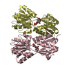
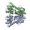
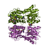
 Structure visualization
 Downloads & links
 Other downloads


 PDBj
PDBj


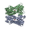
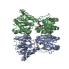
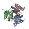
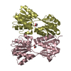
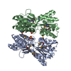
 Assembly
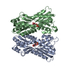
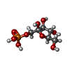
 Type: D-saccharide, beta linking / Mass: 260.136 Da / Num. of mol.: 2
 Mass: 18.015 Da / Num. of mol.: 107 / Source method: isolated from a natural source / Formula: H2O
 Sample preparation
 / Beamline: ID14-2 / Wavelength: 0.933 Å
 Processing