 Movie
Movie Controller

[English] 日本語
 Yorodumi
- PDB-3o23: Human unphosphorylated IGF1-R Kinase domain in complex with an hy... -
+ Open data

- Basic information
| Entry | Database: PDB / ID: 3o23 | ||||||
|---|---|---|---|---|---|---|---|
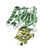
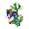
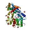
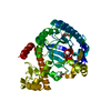
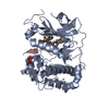
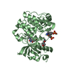
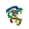
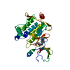
| Title | Human unphosphorylated IGF1-R Kinase domain in complex with an hydantoin inhibitor | ||||||
 Components Components | Insulin-like growth factor 1 receptor | ||||||
 Keywords Keywords | TRANSFERASE/TRANSFERASE INHIBITOR / Protein Kinase / TRANSFERASE-TRANSFERASE INHIBITOR complex | ||||||
| Function / homology |  Function and homology information Function and homology informationinsulin-like growth factor receptor activity / protein kinase complex / insulin-like growth factor binding / Signaling by Type 1 Insulin-like Growth Factor 1 Receptor (IGF1R) / IRS-related events triggered by IGF1R / protein transporter activity / transcytosis / insulin receptor complex / insulin-like growth factor I binding / insulin receptor activity ...insulin-like growth factor receptor activity / protein kinase complex / insulin-like growth factor binding / Signaling by Type 1 Insulin-like Growth Factor 1 Receptor (IGF1R) / IRS-related events triggered by IGF1R / protein transporter activity / transcytosis / insulin receptor complex / insulin-like growth factor I binding / insulin receptor activity / peptidyl-tyrosine autophosphorylation / positive regulation of protein-containing complex disassembly / alphav-beta3 integrin-IGF-1-IGF1R complex / dendritic spine maintenance / regulation of JNK cascade / insulin binding / amyloid-beta clearance / Respiratory syncytial virus (RSV) attachment and entry / insulin receptor substrate binding / SHC-related events triggered by IGF1R / phosphatidylinositol 3-kinase binding / negative regulation of MAPK cascade / insulin-like growth factor receptor signaling pathway / insulin receptor binding / phosphatidylinositol 3-kinase/protein kinase B signal transduction / cellular response to glucose stimulus / receptor protein-tyrosine kinase / cellular response to amyloid-beta / insulin receptor signaling pathway / protein autophosphorylation / positive regulation of cold-induced thermogenesis / protein tyrosine kinase activity / positive regulation of MAPK cascade / positive regulation of phosphatidylinositol 3-kinase/protein kinase B signal transduction / signaling receptor complex / Extra-nuclear estrogen signaling / cilium / immune response / positive regulation of cell migration / axon / positive regulation of cell population proliferation / negative regulation of apoptotic process / nucleolus / signal transduction / ATP binding / membrane / identical protein binding / plasma membrane Similarity search - Function | ||||||
| Biological species |  Homo sapiens (human) Homo sapiens (human) | ||||||
| Method |  X-RAY DIFFRACTION / SYNCHROTRON / MOLECULAR REPLACEMENT / Resolution: 2.1 Å X-RAY DIFFRACTION / SYNCHROTRON / MOLECULAR REPLACEMENT / Resolution: 2.1 Å | ||||||
 Authors Authors | Maignan, S. / Guilloteau, J.P. / Dupuy, A. | ||||||
 Citation Citation | Journal: Bioorg.Med.Chem.Lett. / Year: 2011 Title: Discovery of the first non-ATP competitive IGF-1R kinase inhibitors: Advantages in comparison with competitive inhibitors Authors: Lesuisse, D. / Mauger, J. / Nemecek, C. / Maignan, S. / Boiziau, J. / Harlow, G. / Hittinger, A. / Ruf, S. / Strobel, H. / Nair, A. / Ritter, K. / Malleron, J.L. / Dagallier, A. / El-Ahmad, ...Authors: Lesuisse, D. / Mauger, J. / Nemecek, C. / Maignan, S. / Boiziau, J. / Harlow, G. / Hittinger, A. / Ruf, S. / Strobel, H. / Nair, A. / Ritter, K. / Malleron, J.L. / Dagallier, A. / El-Ahmad, Y. / Guilloteau, J.P. / Guizani, H. / Bouchard, H. / Venot, C. | ||||||
| History |
|
- Structure visualization
| Structure viewer | Molecule: MolmilJmol/JSmol |
|---|
- Downloads & links
-Download
| PDBx/mmCIF format | 3o23.cif.gz | 74.5 KB | Display | PDBx/mmCIF format |
|---|---|---|---|---|
| PDB format | pdb3o23.ent.gz | 54.2 KB | Display | PDB format |
| PDBx/mmJSON format | 3o23.json.gz | Tree view | PDBx/mmJSON format | |
| Others |  Other downloads Other downloads |
-Validation report
| Arichive directory | https://data.pdbj.org/pub/pdb/validation_reports/o2/3o23ftp://data.pdbj.org/pub/pdb/validation_reports/o2/3o23 | HTTPS FTP |
|---|
-Related structure data
| Related structure data |  1jqhS S: Starting model for refinement |
|---|---|
| Similar structure data |


-Links
 PDBj
PDBj


- Assembly
| Deposited unit | 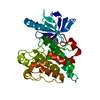
| ||||||||
|---|---|---|---|---|---|---|---|---|---|
| 1 |
| ||||||||
| Unit cell |
|
-Components
| #1: Protein | Mass: 34794.906 Da / Num. of mol.: 1 / Fragment: Kinase domain / Mutation: Y987F Source method: isolated from a genetically manipulated source Source: (gene. exp.) Homo sapiens (human) / Gene: IGF1R / Production host:  References: UniProt: P08069, receptor protein-tyrosine kinase |
|---|---|
| #2: Chemical | ChemComp-MQY / (  Mass: 463.430 Da / Num. of mol.: 1 / Source method: obtained synthetically / Formula: C21H16F3N3O4S Mass: 463.430 Da / Num. of mol.: 1 / Source method: obtained synthetically / Formula: C21H16F3N3O4S |
| #3: Water | ChemComp-HOH /  Mass: 18.015 Da / Num. of mol.: 85 / Source method: isolated from a natural source / Formula: H2O Mass: 18.015 Da / Num. of mol.: 85 / Source method: isolated from a natural source / Formula: H2O |
-Experimental details
-Experiment
| Experiment | Method: X-RAY DIFFRACTION / Number of used crystals: 1 |
|---|
- Sample preparation
| Crystal | Density Matthews: 2.44 Å3/Da / Density % sol: 49.54 % |
|---|---|
| Crystal grow | Temperature: 298 K / Method: vapor diffusion, hanging drop / pH: 6.5 Details: 40% PEG400, 50mM Mes, 100mM NaCl, 10% glycerol, 5mM DTT, pH 6.5, VAPOR DIFFUSION, HANGING DROP, temperature 298.0K |
-Data collection
| Diffraction | Mean temperature: 100 K |
|---|---|
| Diffraction source | Source: SYNCHROTRON / Site: ESRF  / Beamline: ID14-4 / Beamline: ID14-4 |
| Detector | Detector: AREA DETECTOR / Date: Apr 1, 2002 |
| Radiation | Protocol: SINGLE WAVELENGTH / Monochromatic (M) / Laue (L): M / Scattering type: x-ray |
| Radiation wavelength | Relative weight: 1 |
| Reflection | Resolution: 2.1→30 Å / Num. all: 20070 / Num. obs: 18424 / % possible obs: 91.8 % / Observed criterion σ(F): 2 / Observed criterion σ(I): 2 / Rsym value: 0.071 |
- Processing
| Software |
| ||||||||||||||||||||
|---|---|---|---|---|---|---|---|---|---|---|---|---|---|---|---|---|---|---|---|---|---|
| Refinement | Method to determine structure: MOLECULAR REPLACEMENT Starting model: PDB ENTRY 1JQH Resolution: 2.1→30 Å / σ(F): 2 / Stereochemistry target values: Engh & Huber
| ||||||||||||||||||||
| Refinement step | Cycle: LAST / Resolution: 2.1→30 Å
|
