 Movie
Movie Controller

[English] 日本語
 Yorodumi
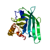
- PDB-3nuf: Crystal structure of a PRD-containing transcription regulator (LS... -
+ Open data

- Basic information
| Entry | Database: PDB / ID: 3nuf | ||||||
|---|---|---|---|---|---|---|---|
| Title | Crystal structure of a PRD-containing transcription regulator (LSEI_2718) from Lactobacillus casei ATCC 334 at 1.38 A resolution | ||||||
 Components Components | PRD-containing transcription regulator | ||||||
 Keywords Keywords | Transcription regulator / STRUCTURAL GENOMICS / JOINT CENTER FOR STRUCTURAL GENOMICS / JCSG / PROTEIN STRUCTURE INITIATIVE / PSI-BIOLOGY / TRANSCRIPTION | ||||||
| Function / homology |  Function and homology information Function and homology information | ||||||
| Biological species |  Lactobacillus casei (bacteria) Lactobacillus casei (bacteria) | ||||||
| Method |  X-RAY DIFFRACTION / SYNCHROTRON / MAD / Resolution: 1.38 Å X-RAY DIFFRACTION / SYNCHROTRON / MAD / Resolution: 1.38 Å | ||||||
 Authors Authors | Joint Center for Structural Genomics (JCSG) | ||||||
 Citation Citation | Journal: To be published Title: Crystal structure of a PRD-containing transcription regulator (LSEI_2718) from Lactobacillus casei ATCC 334 at 1.38 A resolution Authors: Joint Center for Structural Genomics (JCSG) | ||||||
| History |
|
- Structure visualization
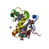
| Structure viewer | Molecule: MolmilJmol/JSmol |
|---|
- Downloads & links
-Download
| PDBx/mmCIF format | 3nuf.cif.gz | 116 KB | Display | PDBx/mmCIF format |
|---|---|---|---|---|
| PDB format | pdb3nuf.ent.gz | 90.8 KB | Display | PDB format |
| PDBx/mmJSON format | 3nuf.json.gz | Tree view | PDBx/mmJSON format | |
| Others |  Other downloads Other downloads |
-Validation report
| Arichive directory | https://data.pdbj.org/pub/pdb/validation_reports/nu/3nufftp://data.pdbj.org/pub/pdb/validation_reports/nu/3nuf | HTTPS FTP |
|---|
-Related structure data
| Similar structure data | |
|---|---|
| Other databases |


-Links
 PDBj
PDBj- Assembly
| Deposited unit | 
| ||||||||
|---|---|---|---|---|---|---|---|---|---|
| 1 | 
| ||||||||
| 2 | 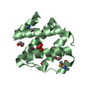
| ||||||||
| 3 |
| ||||||||
| Unit cell |
| ||||||||
| Details | ANALYTICAL SIZE EXCLUSION CHROMATOGRAPHY WITH STATIC LIGHT SCATTERING SUPPORTS THE ASSIGNMENT OF A MONOMER AS A SIGNIFICANT OLIGOMERIZATION STATE IN SOLUTION. ANALYSIS OF THE CRYSTAL PACKING SUGGESTS BOTH THE MONOMER AND THE DIMER IN THE CRYSTAL MIGHT BE STABLE. |
-Components
-Protein , 1 types, 2 molecules AB
| #1: Protein | Mass: 13150.302 Da / Num. of mol.: 2 Source method: isolated from a genetically manipulated source Source: (gene. exp.) Lactobacillus casei (bacteria) / Strain: ATCC 334 / Gene: LSEI_2718 / Plasmid: SpeedET / Production host: |
|---|
-Non-polymers , 5 types, 326 molecules 


| #2: Chemical | ChemComp-EDO /  Mass: 62.068 Da / Num. of mol.: 7 / Source method: obtained synthetically / Formula: C2H6O2 Mass: 62.068 Da / Num. of mol.: 7 / Source method: obtained synthetically / Formula: C2H6O2#3: Chemical | ChemComp-PEG / |  Mass: 106.120 Da / Num. of mol.: 1 / Source method: obtained synthetically / Formula: C4H10O3 Mass: 106.120 Da / Num. of mol.: 1 / Source method: obtained synthetically / Formula: C4H10O3#4: Chemical | ChemComp-SO4 / |  Mass: 96.063 Da / Num. of mol.: 1 / Source method: obtained synthetically / Formula: SO4 Mass: 96.063 Da / Num. of mol.: 1 / Source method: obtained synthetically / Formula: SO4#5: Chemical | ChemComp-PG4 / |  Mass: 194.226 Da / Num. of mol.: 1 / Source method: obtained synthetically / Formula: C8H18O5 / Comment: precipitant*YM Mass: 194.226 Da / Num. of mol.: 1 / Source method: obtained synthetically / Formula: C8H18O5 / Comment: precipitant*YM#6: Water | ChemComp-HOH / | Mass: 18.015 Da / Num. of mol.: 316 / Source method: isolated from a natural source / Formula: H2O |
|---|
-Details
| Has protein modification | Y |
|---|---|
| Sequence details | THE CONSTRUCT WAS EXPRESSED WITH A PURIFICATION TAG MGSDKIHHHHHHENLYFQG. THE TAG WAS REMOVED WITH ...THE CONSTRUCT WAS EXPRESSED WITH A PURIFICATI |
-Experimental details
-Experiment
| Experiment | Method: X-RAY DIFFRACTION / Number of used crystals: 1 |
|---|
- Sample preparation
| Crystal | Density Matthews: 2.14 Å3/Da / Density % sol: 42.47 % Description: DATA WERE SCALED USING XSCALE WITH FRIEDEL PAIRS KEPT AS SEPARATE WHEN COMPUTING R-SYM, COMPLETENESS AND Crystal grow | Temperature: 277 K / Method: vapor diffusion, sitting drop / pH: 7.5 | Details: 1.6000M ammonium sulfate, 4.0000% polyethylene glycol 400, 0.1M HEPES pH 7.5, NANODROP, VAPOR DIFFUSION, SITTING DROP, temperature 277K |
|---|
-Data collection
| Diffraction | Mean temperature: 100 K | |||||||||||||||||||||||||||||||||||||||||||||||||||||||||||||||||||||||||||||
|---|---|---|---|---|---|---|---|---|---|---|---|---|---|---|---|---|---|---|---|---|---|---|---|---|---|---|---|---|---|---|---|---|---|---|---|---|---|---|---|---|---|---|---|---|---|---|---|---|---|---|---|---|---|---|---|---|---|---|---|---|---|---|---|---|---|---|---|---|---|---|---|---|---|---|---|---|---|---|
| Diffraction source | Source: SYNCHROTRON / Site: SSRL  / Beamline: BL9-2 / Wavelength: 0.91837,0.97954,0.97939 / Beamline: BL9-2 / Wavelength: 0.91837,0.97954,0.97939 | |||||||||||||||||||||||||||||||||||||||||||||||||||||||||||||||||||||||||||||
| Detector | Type: MARMOSAIC 325 mm CCD / Detector: CCD / Date: Nov 5, 2009 / Details: Flat collimating mirror, toroid focusing mirror | |||||||||||||||||||||||||||||||||||||||||||||||||||||||||||||||||||||||||||||
| Radiation | Monochromator: Double crystal monochromator / Protocol: MAD / Monochromatic (M) / Laue (L): M / Scattering type: x-ray | |||||||||||||||||||||||||||||||||||||||||||||||||||||||||||||||||||||||||||||
| Radiation wavelength |
| |||||||||||||||||||||||||||||||||||||||||||||||||||||||||||||||||||||||||||||
| Reflection | Resolution: 1.38→28.759 Å / Num. obs: 47575 / % possible obs: 99.4 % / Observed criterion σ(I): -3 / Biso Wilson estimate: 16.639 Å2 / Rmerge(I) obs: 0.054 / Net I/σ(I): 13.65 | |||||||||||||||||||||||||||||||||||||||||||||||||||||||||||||||||||||||||||||
| Reflection shell |
|
-Phasing
| Phasing | Method: MAD |
|---|
- Processing
| Software |
| |||||||||||||||||||||||||||||||||||||||||||||||||||||||||||||||||||||||||||||||||||||
|---|---|---|---|---|---|---|---|---|---|---|---|---|---|---|---|---|---|---|---|---|---|---|---|---|---|---|---|---|---|---|---|---|---|---|---|---|---|---|---|---|---|---|---|---|---|---|---|---|---|---|---|---|---|---|---|---|---|---|---|---|---|---|---|---|---|---|---|---|---|---|---|---|---|---|---|---|---|---|---|---|---|---|---|---|---|---|
| Refinement | Method to determine structure: MAD / Resolution: 1.38→28.759 Å / Cor.coef. Fo:Fc: 0.968 / Cor.coef. Fo:Fc free: 0.958 / Occupancy max: 1 / Occupancy min: 0.25 / SU B: 1.815 / SU ML: 0.037 / Cross valid method: THROUGHOUT / σ(F): 0 / ESU R: 0.061 / ESU R Free: 0.061 Stereochemistry target values: MAXIMUM LIKELIHOOD WITH PHASES Details: 1. HYDROGENS HAVE BEEN ADDED IN THE RIDING POSITIONS. 2. A MET-INHIBITION PROTOCOL WAS USED FOR SELENOMETHIONINE INCORPORATION DURING PROTEIN EXPRESSION. THE OCCUPANCY OF THE SE ATOMS IN THE ...Details: 1. HYDROGENS HAVE BEEN ADDED IN THE RIDING POSITIONS. 2. A MET-INHIBITION PROTOCOL WAS USED FOR SELENOMETHIONINE INCORPORATION DURING PROTEIN EXPRESSION. THE OCCUPANCY OF THE SE ATOMS IN THE MSE RESIDUES WAS REDUCED TO 0.75 FOR THE REDUCED SCATTERING POWER DUE TO PARTIAL S-MET INCORPORATION. 3. ATOM RECORDS CONTAIN SUM OF TLS AND RESIDUAL B FACTORS. ANISOU RECORDS CONTAIN SUM OF TLS AND RESIDUAL U FACTORS. 4. WATERS WERE EXCLUDED FROM AUTOMATIC TLS ASSIGNMENT. 5. SULFATE (SO4) IONS, AND PEG-400 FRAGMENTS (PG4 AND PEG) ARE MODELED FROM CRYSTALLIZATION CONDITIONS. 6. ETHYLENE GLYCOL (EDO) MOLECULES ARE MODELED FROM CRYO CONDITIONS. 7. THERE IS A 5.4 SIGMA PEAK IN THE FOFC ELECTRON DENSITY MAP NEAR CARBONYL OXYGEN OF VALINE 106, WHICH COULD BE A WATER OR AMMONIUM ION, THIS REMAINS UNMODELLED.
| |||||||||||||||||||||||||||||||||||||||||||||||||||||||||||||||||||||||||||||||||||||
| Solvent computation | Ion probe radii: 0.8 Å / Shrinkage radii: 0.8 Å / VDW probe radii: 1.2 Å / Solvent model: MASK | |||||||||||||||||||||||||||||||||||||||||||||||||||||||||||||||||||||||||||||||||||||
| Displacement parameters | Biso max: 64.26 Å2 / Biso mean: 20.531 Å2 / Biso min: 3.4 Å2
| |||||||||||||||||||||||||||||||||||||||||||||||||||||||||||||||||||||||||||||||||||||
| Refinement step | Cycle: LAST / Resolution: 1.38→28.759 Å
| |||||||||||||||||||||||||||||||||||||||||||||||||||||||||||||||||||||||||||||||||||||
| Refine LS restraints |
| |||||||||||||||||||||||||||||||||||||||||||||||||||||||||||||||||||||||||||||||||||||
| LS refinement shell | Resolution: 1.38→1.415 Å / Total num. of bins used: 20
| |||||||||||||||||||||||||||||||||||||||||||||||||||||||||||||||||||||||||||||||||||||
| Refinement TLS params. | Method: refined / Refine-ID: X-RAY DIFFRACTION
| |||||||||||||||||||||||||||||||||||||||||||||||||||||||||||||||||||||||||||||||||||||
| Refinement TLS group |
|
