- PDB-3nsg: Crystal Structure of OmpF, an Outer Membrane Protein from Salmone... -
+
Open data
ID or keywords:
Loading...
-
Basic information
Entry
Database: PDB / ID: 3nsg
Title
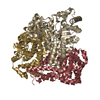
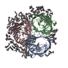
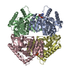
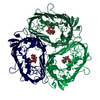
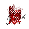
Crystal Structure of OmpF, an Outer Membrane Protein from Salmonella typhi
Components
Outer membrane protein F
Keywords
MEMBRANE PROTEIN / Porin / beta barrel / outer membrane protein / Beta Barrel Membrane protein
Function / homology
Function and homology information
porin activity / pore complex / cell outer membrane / monoatomic ion transmembrane transport Similarity search - Function
Porin, gammaproteobacterial / Porin, Gram-negative type, conserved site / General diffusion Gram-negative porins signature. / Porin domain, Gram-negative type / Gram-negative porin / Porin / Porin, Gram-negative type / : / Porin domain superfamily / Porin ...Porin, gammaproteobacterial / Porin, Gram-negative type, conserved site / General diffusion Gram-negative porins signature. / Porin domain, Gram-negative type / Gram-negative porin / Porin / Porin, Gram-negative type / : / Porin domain superfamily / Porin / Beta Barrel / Mainly Beta Similarity search - Domain/homology
Mass: 18.015 Da / Num. of mol.: 387 / Source method: isolated from a natural source / Formula: H2O
-
Details
Has protein modification
Y
Sequence details
A SEQUENCE DATABASE REFERENCE FOR THIS PROTEIN WHCIH HAS DERIVED FROM SALMONELLA TYPHI DOES NOT ...A SEQUENCE DATABASE REFERENCE FOR THIS PROTEIN WHCIH HAS DERIVED FROM SALMONELLA TYPHI DOES NOT CURRENTLY EXIST.
-
Experimental details
-
Experiment
Experiment
Method: X-RAY DIFFRACTION / Number of used crystals: 1
-
Sample preparation
Crystal
Density Matthews: 3.9 Å3/Da / Density % sol: 68.48 % / Mosaicity: 0.721 °
Crystal grow
Method: vapor diffusion, hanging drop / pH: 5.4 Details: 0.1M Tri sodium citrate, 0.25M potassium sodium tartrate, 2M Ammonium sulphate, pH 5.4, VAPOR DIFFUSION, HANGING DROP
Resolution: 2.79→50 Å / Num. obs: 38061 / % possible obs: 84.4 % / Redundancy: 7.7 % / Rmerge(I) obs: 0.092 / Χ2: 1.116 / Net I/σ(I): 8
Reflection shell
Resolution (Å)
Redundancy (%)
Rmerge(I) obs
Num. unique all
Χ2
Diffraction-ID
% possible all
2.8-2.9
4.8
0.364
2529
0.63
1
57.1
2.9-3.02
5.4
0.32
2886
0.662
1
64.8
3.02-3.15
5.9
0.244
3198
0.649
1
71.8
3.15-3.32
6.5
0.206
3361
0.661
1
75.7
3.32-3.53
7.2
0.151
3712
0.718
1
82.8
3.53-3.8
7.7
0.119
4107
0.802
1
91.9
3.8-4.18
8.5
0.085
4433
0.915
1
98.6
4.18-4.79
9.3
0.071
4527
1.181
1
99.8
4.79-6.03
9.4
0.084
4561
1.466
1
99.9
6.03-50
9
0.055
4747
2.029
1
99.5
-
Processing
Software
Name
Version
Classification
NB
DENZO
datareduction
SCALEPACK
datascaling
REFMAC
5.5.0072
refinement
PDB_EXTRACT
3.1
dataextraction
Refinement
Method to determine structure: SAD / Resolution: 2.79→42.59 Å / Cor.coef. Fo:Fc: 0.891 / Cor.coef. Fo:Fc free: 0.801 / Occupancy max: 1 / Occupancy min: 0.5 / SU B: 24.658 / SU ML: 0.461 / Cross valid method: THROUGHOUT / ESU R Free: 0.518 / Stereochemistry target values: MAXIMUM LIKELIHOOD Details: HYDROGENS HAVE BEEN ADDED IN THE RIDING POSITIONS U VALUES: REFINED INDIVIDUALLY
Rfactor
Num. reflection
% reflection
Selection details
Rfree
0.33672
1898
5 %
RANDOM
Rwork
0.25489
-
-
-
obs
0.25887
36118
84.08 %
-
Solvent computation
Ion probe radii: 0.8 Å / Shrinkage radii: 0.8 Å / VDW probe radii: 1.4 Å / Solvent model: MASK
In the structure databanks used in Yorodumi, some data are registered as the other names, "COVID-19 virus" and "2019-nCoV". Here are the details of the virus and the list of structure data.
Jan 31, 2019. EMDB accession codes are about to change! (news from PDBe EMDB page)
EMDB accession codes are about to change! (news from PDBe EMDB page)
The allocation of 4 digits for EMDB accession codes will soon come to an end. Whilst these codes will remain in use, new EMDB accession codes will include an additional digit and will expand incrementally as the available range of codes is exhausted. The current 4-digit format prefixed with “EMD-” (i.e. EMD-XXXX) will advance to a 5-digit format (i.e. EMD-XXXXX), and so on. It is currently estimated that the 4-digit codes will be depleted around Spring 2019, at which point the 5-digit format will come into force.
The EM Navigator/Yorodumi systems omit the EMD- prefix.
Related info.:Q: What is EMD? / ID/Accession-code notation in Yorodumi/EM Navigator
Yorodumi is a browser for structure data from EMDB, PDB, SASBDB, etc.
This page is also the successor to EM Navigator detail page, and also detail information page/front-end page for Omokage search.
The word "yorodu" (or yorozu) is an old Japanese word meaning "ten thousand". "mi" (miru) is to see.
Related info.:EMDB / PDB / SASBDB / Comparison of 3 databanks / Yorodumi Search / Aug 31, 2016. New EM Navigator & Yorodumi / Yorodumi Papers / Jmol/JSmol / Function and homology information / Changes in new EM Navigator and Yorodumi
 Movie
Movie Controller

 Yorodumi
 Open data
 Basic information
 Components
 Keywords
 Function and homology information
 Salmonella enterica subsp. enterica serovar Typhi (bacteria)
 X-RAY DIFFRACTION /
  Authors
 Citation
 Structure visualization
 Downloads & links
 Other downloads


 PDBj
PDBj


 Assembly
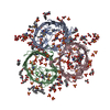


 Mass: 96.063 Da / Num. of mol.: 80 / Source method: obtained synthetically / Formula: SO4
 Mass: 92.094 Da / Num. of mol.: 38 / Source method: obtained synthetically / Formula: C3H8O3
 Mass: 229.402 Da / Num. of mol.: 17 / Source method: obtained synthetically / Formula: C14H31NO / Comment: LDAO, detergent*YM
 Mass: 163.215 Da / Num. of mol.: 6 / Source method: obtained synthetically / Formula: C7H17NO3 / Comment: pH buffer*YM
 Mass: 189.100 Da / Num. of mol.: 4 / Source method: obtained synthetically / Formula: C6H5O7
 Mass: 150.087 Da / Num. of mol.: 2 / Source method: obtained synthetically / Formula: C4H6O6
 Sample preparation
 / Beamline: BM14 / Wavelength: 0.979 Å
 Processing