 Movie
Movie Controller

[English] 日本語
 Yorodumi
- PDB-3nq0: Crystal Structure of Tyrosinase from Bacillus megaterium crystall... -
+ Open data

- Basic information
| Entry | Database: PDB / ID: 3nq0 | ||||||
|---|---|---|---|---|---|---|---|
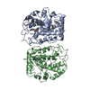
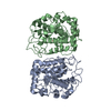
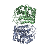
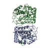
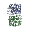
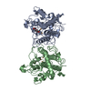
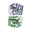
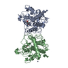
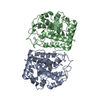
| Title | Crystal Structure of Tyrosinase from Bacillus megaterium crystallized in the absence of Zinc | ||||||
 Components Components | Tyrosinase | ||||||
 Keywords Keywords | OXIDOREDUCTASE / tyrosinase / type3 copper proteins / Bacillus megaterium | ||||||
| Function / homology |  Function and homology information Function and homology information | ||||||
| Biological species |  Bacillus megaterium (bacteria) Bacillus megaterium (bacteria) | ||||||
| Method |  X-RAY DIFFRACTION / SYNCHROTRON / MOLECULAR REPLACEMENT / molecular replacement / Resolution: 2.2 Å X-RAY DIFFRACTION / SYNCHROTRON / MOLECULAR REPLACEMENT / molecular replacement / Resolution: 2.2 Å | ||||||
 Authors Authors | Sendovski, M. / Kanteev, M. / Adir, N. / Fishman, A. | ||||||
 Citation Citation | Journal: J.Mol.Biol. / Year: 2011 Title: First structures of an active bacterial tyrosinase reveal copper plasticity. Authors: Sendovski, M. / Kanteev, M. / Shuster Ben-Yosef, V. / Adir, N. / Fishman, A. #1: Journal: To be PublishedTitle: Crystallization and preliminary x-ray crystallographic analysis of a bacterial tyrosinase from Bacillus megaterium Authors: Sendovski, M. / Kanteev, M. / Adir, N. / Fishman, A. | ||||||
| History |
|
- Structure visualization
| Structure viewer | Molecule: MolmilJmol/JSmol |
|---|
- Downloads & links
-Download
| PDBx/mmCIF format | 3nq0.cif.gz | 127.7 KB | Display | PDBx/mmCIF format |
|---|---|---|---|---|
| PDB format | pdb3nq0.ent.gz | 98.2 KB | Display | PDB format |
| PDBx/mmJSON format | 3nq0.json.gz | Tree view | PDBx/mmJSON format | |
| Others |  Other downloads Other downloads |
-Validation report
| Arichive directory | https://data.pdbj.org/pub/pdb/validation_reports/nq/3nq0ftp://data.pdbj.org/pub/pdb/validation_reports/nq/3nq0 | HTTPS FTP |
|---|
-Related structure data
| Related structure data |  3nm8SC  3npyC  3nq1C  3nq5C  3ntmC S: Starting model for refinement C: citing same article ( |
|---|---|
| Similar structure data |


-Links
 PDBj
PDBj- Assembly
| Deposited unit | 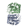
| ||||||||
|---|---|---|---|---|---|---|---|---|---|
| 1 |
| ||||||||
| Unit cell |
|
-Components
| #1: Protein | Mass: 35284.500 Da / Num. of mol.: 2 Source method: isolated from a genetically manipulated source Source: (gene. exp.) Bacillus megaterium (bacteria) / Plasmid: pET9d / Production host: #2: Chemical |   Mass: 63.546 Da / Num. of mol.: 2 / Source method: obtained synthetically / Formula: Cu Mass: 63.546 Da / Num. of mol.: 2 / Source method: obtained synthetically / Formula: Cu#3: Water | ChemComp-HOH / |  Mass: 18.015 Da / Num. of mol.: 159 / Source method: isolated from a natural source / Formula: H2O Mass: 18.015 Da / Num. of mol.: 159 / Source method: isolated from a natural source / Formula: H2O |
|---|
-Experimental details
-Experiment
| Experiment | Method: X-RAY DIFFRACTION / Number of used crystals: 1 |
|---|
- Sample preparation
| Crystal | Density Matthews: 2.2 Å3/Da / Density % sol: 44.12 % |
|---|---|
| Crystal grow | Temperature: 293 K / Method: vapor diffusion, hanging drop / pH: 6.5 Details: PEG 8000, sodium cacodylate, pH 6.5, vapor diffusion, hanging drop, temperature 293K |
-Data collection
| Diffraction | Mean temperature: 100 K | |||||||||||||||||||||||||||||||||||||||||||||||||||||||||||||||||||||||||||||
|---|---|---|---|---|---|---|---|---|---|---|---|---|---|---|---|---|---|---|---|---|---|---|---|---|---|---|---|---|---|---|---|---|---|---|---|---|---|---|---|---|---|---|---|---|---|---|---|---|---|---|---|---|---|---|---|---|---|---|---|---|---|---|---|---|---|---|---|---|---|---|---|---|---|---|---|---|---|---|
| Diffraction source | Source: SYNCHROTRON / Site: ESRF  / Beamline: ID14-1 / Wavelength: 0.934 Å / Beamline: ID14-1 / Wavelength: 0.934 Å | |||||||||||||||||||||||||||||||||||||||||||||||||||||||||||||||||||||||||||||
| Detector | Type: ADSC QUANTUM 210 / Detector: CCD / Date: Mar 5, 2010 | |||||||||||||||||||||||||||||||||||||||||||||||||||||||||||||||||||||||||||||
| Radiation | Protocol: SINGLE WAVELENGTH / Monochromatic (M) / Laue (L): M / Scattering type: x-ray | |||||||||||||||||||||||||||||||||||||||||||||||||||||||||||||||||||||||||||||
| Radiation wavelength | Wavelength: 0.934 Å / Relative weight: 1 | |||||||||||||||||||||||||||||||||||||||||||||||||||||||||||||||||||||||||||||
| Reflection | Resolution: 2.2→82.551 Å / Num. all: 31201 / Num. obs: 29606 / % possible obs: 95.1 % / Redundancy: 2.1 % / Biso Wilson estimate: 12.4 Å2 / Rsym value: 0.1 / Net I/σ(I): 8.3 | |||||||||||||||||||||||||||||||||||||||||||||||||||||||||||||||||||||||||||||
| Reflection shell |
|
-Phasing
| Phasing | Method: molecular replacement | |||||||||
|---|---|---|---|---|---|---|---|---|---|---|
| Phasing MR | Rfactor: 37.64 / Model details: Phaser MODE: MR_AUTO
|
- Processing
| Software |
| ||||||||||||||||||||||||||||||||
|---|---|---|---|---|---|---|---|---|---|---|---|---|---|---|---|---|---|---|---|---|---|---|---|---|---|---|---|---|---|---|---|---|---|
| Refinement | Method to determine structure: MOLECULAR REPLACEMENT Starting model: PDB ENTRY 3NM8 Resolution: 2.2→41.275 Å / Occupancy max: 1 / Occupancy min: 1 / SU ML: 0.36 / σ(F): 0.02 / Stereochemistry target values: ML
| ||||||||||||||||||||||||||||||||
| Solvent computation | Shrinkage radii: 0.9 Å / VDW probe radii: 1.11 Å / Solvent model: FLAT BULK SOLVENT MODEL / Bsol: 40.351 Å2 / ksol: 0.4 e/Å3 | ||||||||||||||||||||||||||||||||
| Displacement parameters | Biso mean: 17.4718 Å2
| ||||||||||||||||||||||||||||||||
| Refine analyze | Luzzati coordinate error obs: 0.36 Å / Luzzati sigma a obs: 0.38 Å | ||||||||||||||||||||||||||||||||
| Refinement step | Cycle: LAST / Resolution: 2.2→41.275 Å
| ||||||||||||||||||||||||||||||||
| Refine LS restraints |
|
