transition metal ion binding / periplasmic space / electron transfer activity / copper ion binding / zinc ion binding / identical protein binding / plasma membrane Similarity search - Function
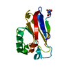
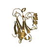
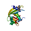
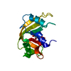
Azurin / : / Blue (type 1) copper domain / Copper binding proteins, plastocyanin/azurin family / Blue (type 1) copper protein, binding site / Type-1 copper (blue) proteins signature. / Cupredoxins - blue copper proteins / Cupredoxin / Immunoglobulin-like / Sandwich / Mainly Beta Similarity search - Domain/homology
Mass: 18.015 Da / Num. of mol.: 60 / Source method: isolated from a natural source / Formula: H2O
Has protein modification
Y
-
Experimental details
-
Experiment
Experiment
Method: X-RAY DIFFRACTION / Number of used crystals: 1
-
Sample preparation
Crystal
Density Matthews: 2.25 Å3/Da / Density % sol: 45.44 %
Crystal grow
Temperature: 298 K / Method: vapor diffusion, sitting drop / pH: 7 Details: 25-30% PEG 4000 100 mM LiNO3 20 mM CuCl2 100 mM Tris pH 7, VAPOR DIFFUSION, SITTING DROP, temperature 298K
-
Data collection
Diffraction
Mean temperature: 100 K
Diffraction source
Source: ROTATING ANODE / Type: RIGAKU / Wavelength: 1.54 Å
Resolution: 2.25→33.923 Å / Cor.coef. Fo:Fc: 0.944 / Cor.coef. Fo:Fc free: 0.902 / SU B: 0.008 / SU ML: 0 / Cross valid method: THROUGHOUT / ESU R Free: 0.278 / Stereochemistry target values: MAXIMUM LIKELIHOOD / Details: HYDROGENS HAVE BEEN ADDED IN THE RIDING POSITIONS
Rfactor
Num. reflection
% reflection
Selection details
Rfree
0.27616
289
4.9 %
RANDOM
Rwork
0.21621
-
-
-
obs
0.21908
5614
94.07 %
-
Solvent computation
Ion probe radii: 0.8 Å / Shrinkage radii: 0.8 Å / VDW probe radii: 1.4 Å / Solvent model: MASK
Displacement parameters
Biso mean: 43.827 Å2
Baniso -1
Baniso -2
Baniso -3
1-
-0.07 Å2
0 Å2
0 Å2
2-
-
0.06 Å2
0 Å2
3-
-
-
0.01 Å2
Refinement step
Cycle: LAST / Resolution: 2.25→33.923 Å
Protein
Nucleic acid
Ligand
Solvent
Total
Num. atoms
976
0
10
60
1046
LS refinement shell
Resolution: 2.25→2.308 Å / Total num. of bins used: 20
Rfactor
Num. reflection
% reflection
Rfree
0.225
18
-
Rwork
0.217
404
-
obs
-
404
89.79 %
+
About Yorodumi
-
News
-
Feb 9, 2022. New format data for meta-information of EMDB entries
New format data for meta-information of EMDB entries
Version 3 of the EMDB header file is now the official format.
The previous official version 1.9 will be removed from the archive.
In the structure databanks used in Yorodumi, some data are registered as the other names, "COVID-19 virus" and "2019-nCoV". Here are the details of the virus and the list of structure data.
Jan 31, 2019. EMDB accession codes are about to change! (news from PDBe EMDB page)
EMDB accession codes are about to change! (news from PDBe EMDB page)
The allocation of 4 digits for EMDB accession codes will soon come to an end. Whilst these codes will remain in use, new EMDB accession codes will include an additional digit and will expand incrementally as the available range of codes is exhausted. The current 4-digit format prefixed with “EMD-” (i.e. EMD-XXXX) will advance to a 5-digit format (i.e. EMD-XXXXX), and so on. It is currently estimated that the 4-digit codes will be depleted around Spring 2019, at which point the 5-digit format will come into force.
The EM Navigator/Yorodumi systems omit the EMD- prefix.
Related info.:Q: What is EMD? / ID/Accession-code notation in Yorodumi/EM Navigator
Yorodumi is a browser for structure data from EMDB, PDB, SASBDB, etc.
This page is also the successor to EM Navigator detail page, and also detail information page/front-end page for Omokage search.
The word "yorodu" (or yorozu) is an old Japanese word meaning "ten thousand". "mi" (miru) is to see.
Related info.:EMDB / PDB / SASBDB / Comparison of 3 databanks / Yorodumi Search / Aug 31, 2016. New EM Navigator & Yorodumi / Yorodumi Papers / Jmol/JSmol / Function and homology information / Changes in new EM Navigator and Yorodumi
 Movie
Movie Controller


 Open data
 Basic information
 Components
 Keywords
 Function and homology information
 Pseudomonas aeruginosa (bacteria)
 X-RAY DIFFRACTION /
  Authors
 Citation
 Structure visualization
 Downloads & links
 Other downloads


 PDBj
PDBj Assembly
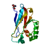

 Mass: 63.546 Da / Num. of mol.: 2 / Source method: obtained synthetically / Formula: Cu
 Mass: 122.143 Da / Num. of mol.: 1 / Source method: obtained synthetically / Formula: C4H12NO3 / Comment: pH buffer*YM
 Mass: 18.015 Da / Num. of mol.: 60 / Source method: isolated from a natural source / Formula: H2O
 Sample preparation
 Processing