Mass: 18.015 Da / Num. of mol.: 368 / Source method: isolated from a natural source / Formula: H2O
Has protein modification
Y
Sequence details
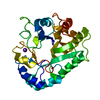
THE CONSTRUCT (20-294) WAS EXPRESSED WITH A PURIFICATION TAG MGSDKIHHHHHHENLYFQG. THE TAG WAS ...THE CONSTRUCT (20-294) WAS EXPRESSED WITH A PURIFICATION TAG MGSDKIHHHHHHENLYFQG. THE TAG WAS REMOVED WITH TEV PROTEASE LEAVING ONLY A GLYCINE (0) FOLLOWED BY THE TARGET SEQUENCE.
-
Experimental details
-
Experiment
Experiment
Method: X-RAY DIFFRACTION / Number of used crystals: 1
-
Sample preparation
Crystal
Density Matthews: 2.54 Å3/Da / Density % sol: 51.48 %
Crystal grow
Temperature: 277 K / Method: vapor diffusion, sitting drop / pH: 5.5 Details: 40.000000000% PEG-600, 0.1M Citrate pH 5.5, VAPOR DIFFUSION,SITTING DROP,NANODROP, temperature 277K, VAPOR DIFFUSION, SITTING DROP
Monochromator: Single crystal Si(111) bent monochromator (horizontal focusing) Protocol: MAD / Monochromatic (M) / Laue (L): M / Scattering type: x-ray
Radiation wavelength
ID
Wavelength (Å)
Relative weight
1
0.91837
1
2
0.97941
1
3
0.97894
1
Reflection
Resolution: 1.35→27.993 Å / Num. obs: 68533 / % possible obs: 97.7 % / Observed criterion σ(I): -3 / Biso Wilson estimate: 10.556 Å2 / Rmerge F obs: 0.164 / Rmerge(I) obs: 0.058 / Rrim(I) all: 0.075 / Net I/σ(I): 10.61 / Num. measured all: 297194
Reflection shell
Resolution (Å)
Highest resolution (Å)
Rmerge F obs
Rmerge(I) obs
Mean I/σ(I) obs
Num. measured obs
Num. possible
Num. unique obs
Rrim(I) all
% possible all
1.35-1.4
0.861
0.505
1.9
28471
13740
13087
0.657
95.2
1.4-1.45
0.684
0.396
2.4
25252
11841
11410
0.516
96.4
1.45-1.52
0.531
0.308
3.1
30507
14080
13642
0.401
96.9
1.52-1.6
0.373
0.222
4.2
29318
13271
12963
0.288
97.7
1.6-1.7
0.292
0.168
5.5
29515
13174
12921
0.219
98.1
1.7-1.83
0.197
0.118
7.6
30053
13197
12944
0.153
98.1
1.83-2.02
0.124
0.075
11.5
31493
13629
13432
0.098
98.6
2.02-2.31
0.075
0.05
16.5
30508
13094
12930
0.065
98.7
2.31-2.91
0.052
0.038
21
30934
13209
13082
0.049
99
2.91
0.027
0.025
31.6
31143
13253
13079
0.032
98.7
-
Phasing
Phasing
Method: MAD
-
Processing
Software
Name
Version
Classification
NB
SHELX
phasing
REFMAC
5.5.0110
refinement
XSCALE
datascaling
PDB_EXTRACT
3.1
dataextraction
XDS
datareduction
SHELXD
phasing
autoSHARP
phasing
Refinement
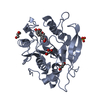
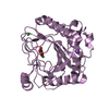
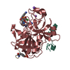
Method to determine structure: MAD / Resolution: 1.35→27.993 Å / Cor.coef. Fo:Fc: 0.972 / Cor.coef. Fo:Fc free: 0.967 / Occupancy max: 1 / Occupancy min: 0.11 / SU B: 1.292 / SU ML: 0.027 / Cross valid method: THROUGHOUT / σ(F): 0 / ESU R: 0.046 / ESU R Free: 0.045 Stereochemistry target values: MAXIMUM LIKELIHOOD WITH PHASES Details: (1). HYDROGENS HAVE BEEN ADDED IN THE RIDING POSITIONS. (2). ATOM RECORD CONTAINS SUM OF TLS AND RESIDUAL B FACTORS. ANISOU RECORD CONTAINS SUM OF TLS AND RESIDUAL U FACTORS. (3). A MET- ...Details: (1). HYDROGENS HAVE BEEN ADDED IN THE RIDING POSITIONS. (2). ATOM RECORD CONTAINS SUM OF TLS AND RESIDUAL B FACTORS. ANISOU RECORD CONTAINS SUM OF TLS AND RESIDUAL U FACTORS. (3). A MET-INHIBITION PROTOCOL WAS USED FOR SELENOMETHIONINE INCORPORATION DURING PROTEIN EXPRESSION. THE OCCUPANCY OF THE SE ATOMS IN THE MSE RESIDUES WAS REDUCED TO 0.75 TO ACCOUNT FOR THE REDUCED SCATTERING POWER DUE TO PARTIAL S-MET INCORPORATION. (4). CL AND CITRATE (CIT) IONS, AND PEG-600 FRAGMENTS (PEG) FROM CRYSTALLIZATION CONDITION WERE MODELED INTO THE STRUCTURE. (5). RAMACHANDRAN OUTLIER OF RESIDUE GLN144 IS SUPPORTED BY ELECTRON DENSITY.
Rfactor
Num. reflection
% reflection
Selection details
Rfree
0.1595
3466
5.1 %
RANDOM
Rwork
0.1468
65006
-
-
obs
0.1475
68472
98.8 %
-
Solvent computation
Ion probe radii: 0.8 Å / Shrinkage radii: 0.8 Å / VDW probe radii: 1.4 Å / Solvent model: MASK
In the structure databanks used in Yorodumi, some data are registered as the other names, "COVID-19 virus" and "2019-nCoV". Here are the details of the virus and the list of structure data.
Jan 31, 2019. EMDB accession codes are about to change! (news from PDBe EMDB page)
EMDB accession codes are about to change! (news from PDBe EMDB page)
The allocation of 4 digits for EMDB accession codes will soon come to an end. Whilst these codes will remain in use, new EMDB accession codes will include an additional digit and will expand incrementally as the available range of codes is exhausted. The current 4-digit format prefixed with “EMD-” (i.e. EMD-XXXX) will advance to a 5-digit format (i.e. EMD-XXXXX), and so on. It is currently estimated that the 4-digit codes will be depleted around Spring 2019, at which point the 5-digit format will come into force.
The EM Navigator/Yorodumi systems omit the EMD- prefix.
Related info.:Q: What is EMD? / ID/Accession-code notation in Yorodumi/EM Navigator
Yorodumi is a browser for structure data from EMDB, PDB, SASBDB, etc.
This page is also the successor to EM Navigator detail page, and also detail information page/front-end page for Omokage search.
The word "yorodu" (or yorozu) is an old Japanese word meaning "ten thousand". "mi" (miru) is to see.
Related info.:EMDB / PDB / SASBDB / Comparison of 3 databanks / Yorodumi Search / Aug 31, 2016. New EM Navigator & Yorodumi / Yorodumi Papers / Jmol/JSmol / Function and homology information / Changes in new EM Navigator and Yorodumi
 Movie
Movie Controller

 Yorodumi
 Open data
 Basic information
 Components
 Keywords
 Function and homology information
 Bacteroides caccae (bacteria)
 X-RAY DIFFRACTION /
  Authors
 Citation
 Structure visualization
 Downloads & links
 Other downloads
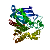

 PDBj
PDBj Assembly
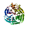

 Mass: 35.453 Da / Num. of mol.: 1 / Source method: obtained synthetically / Formula: Cl
 Mass: 192.124 Da / Num. of mol.: 1 / Source method: obtained synthetically / Formula: C6H8O7
 Mass: 106.120 Da / Num. of mol.: 2 / Source method: obtained synthetically / Formula: C4H10O3
 Mass: 18.015 Da / Num. of mol.: 368 / Source method: isolated from a natural source / Formula: H2O
 Sample preparation
 / Beamline: BL11-1 / Wavelength: 0.91837,0.97941,0.97894
 Processing