 Movie
Movie Controller

[English] 日本語
 Yorodumi
- PDB-3nnt: Crystal Structure of K170M Mutant of Type I 3-Dehydroquinate Dehy... -
+ Open data

- Basic information
| Entry | Database: PDB / ID: 3nnt | ||||||
|---|---|---|---|---|---|---|---|
| Title | Crystal Structure of K170M Mutant of Type I 3-Dehydroquinate Dehydratase (aroD) from Salmonella typhimurium LT2 in Non-Covalent Complex with Dehydroquinate. | ||||||
 Components Components | 3-dehydroquinate dehydratase | ||||||
 Keywords Keywords | LYASE / Structural Genomics / Center for Structural Genomics of Infectious Diseases / CSGID | ||||||
| Function / homology |  Function and homology information Function and homology information3,4-dihydroxybenzoate biosynthetic process / 3-dehydroquinate dehydratase / 3-dehydroquinate dehydratase activity / chorismate biosynthetic process / aromatic amino acid biosynthetic process / amino acid biosynthetic process / cytosol Similarity search - Function | ||||||
| Biological species |  Salmonella enterica subsp. enterica serovar Typhimurium (bacteria) Salmonella enterica subsp. enterica serovar Typhimurium (bacteria) | ||||||
| Method |  X-RAY DIFFRACTION / SYNCHROTRON / MOLECULAR REPLACEMENT / Resolution: 1.6 Å X-RAY DIFFRACTION / SYNCHROTRON / MOLECULAR REPLACEMENT / Resolution: 1.6 Å | ||||||
 Authors Authors | Minasov, G. / Light, S.H. / Shuvalova, L. / Papazisi, L. / Anderson, W.F. / Center for Structural Genomics of Infectious Diseases (CSGID) | ||||||
 Citation Citation | Journal: J.Biol.Chem. / Year: 2011 Title: Insights into the mechanism of type I dehydroquinate dehydratases from structures of reaction intermediates. Authors: Light, S.H. / Minasov, G. / Shuvalova, L. / Duban, M.E. / Caffrey, M. / Anderson, W.F. / Lavie, A. | ||||||
| History |
|
- Structure visualization
| Structure viewer | Molecule: MolmilJmol/JSmol |
|---|
- Downloads & links
-Download
| PDBx/mmCIF format | 3nnt.cif.gz | 128.1 KB | Display | PDBx/mmCIF format |
|---|---|---|---|---|
| PDB format | pdb3nnt.ent.gz | 98.5 KB | Display | PDB format |
| PDBx/mmJSON format | 3nnt.json.gz | Tree view | PDBx/mmJSON format | |
| Others |  Other downloads Other downloads |
-Validation report
| Arichive directory | https://data.pdbj.org/pub/pdb/validation_reports/nn/3nntftp://data.pdbj.org/pub/pdb/validation_reports/nn/3nnt | HTTPS FTP |
|---|
-Related structure data
| Related structure data |  3js3C  3m7wC  3lb0S S: Starting model for refinement C: citing same article ( |
|---|---|
| Similar structure data | |
| Other databases |


-Links
 PDBj
PDBj
- Assembly
| Deposited unit | 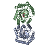
| ||||||||
|---|---|---|---|---|---|---|---|---|---|
| 1 |
| ||||||||
| Unit cell |
|
-Components
| #1: Protein | Mass: 30103.324 Da / Num. of mol.: 2 / Mutation: K170M Source method: isolated from a genetically manipulated source Source: (gene. exp.) Salmonella enterica subsp. enterica serovar Typhimurium (bacteria)Strain: LT2 / Gene: aroD, STM1358 / Plasmid: pMCSG7 / Production host: #2: Chemical |   Mass: 190.151 Da / Num. of mol.: 2 / Source method: obtained synthetically / Formula: C7H10O6 Mass: 190.151 Da / Num. of mol.: 2 / Source method: obtained synthetically / Formula: C7H10O6#3: Water | ChemComp-HOH / |  Mass: 18.015 Da / Num. of mol.: 525 / Source method: isolated from a natural source / Formula: H2O Mass: 18.015 Da / Num. of mol.: 525 / Source method: isolated from a natural source / Formula: H2O |
|---|
-Experimental details
-Experiment
| Experiment | Method: X-RAY DIFFRACTION / Number of used crystals: 1 |
|---|
- Sample preparation
| Crystal | Density Matthews: 1.96 Å3/Da / Density % sol: 37.26 % |
|---|---|
| Crystal grow | Temperature: 295 K / Method: vapor diffusion, sitting drop / pH: 3 Details: Protein solution: 7.5 mG/mL, 0.25M Sodium chloride, 0.01M Tris pH 8.3, 2mM 3-Dehydroquinic acid (DHR); Screen solution: Classics F9, 0.05M Potassium phosphate, 20%(w/v) PEG 8000., VAPOR ...Details: Protein solution: 7.5 mG/mL, 0.25M Sodium chloride, 0.01M Tris pH 8.3, 2mM 3-Dehydroquinic acid (DHR); Screen solution: Classics F9, 0.05M Potassium phosphate, 20%(w/v) PEG 8000., VAPOR DIFFUSION, SITTING DROP, temperature 295K |
-Data collection
| Diffraction | Mean temperature: 100 K |
|---|---|
| Diffraction source | Source: SYNCHROTRON / Site: APS  / Beamline: 21-ID-G / Wavelength: 0.97856 Å / Beamline: 21-ID-G / Wavelength: 0.97856 Å |
| Detector | Type: MARMOSAIC 300 mm CCD / Detector: CCD / Date: Jun 18, 2010 / Details: Beryllium lenses |
| Radiation | Monochromator: Diamond / Protocol: SINGLE WAVELENGTH / Monochromatic (M) / Laue (L): M / Scattering type: x-ray |
| Radiation wavelength | Wavelength: 0.97856 Å / Relative weight: 1 |
| Reflection | Resolution: 1.6→30 Å / Num. all: 58590 / Num. obs: 58590 / % possible obs: 96.7 % / Observed criterion σ(I): -3 / Redundancy: 2 % / Biso Wilson estimate: 24 Å2 / Rmerge(I) obs: 0.03 / Net I/σ(I): 23.2 |
| Reflection shell | Resolution: 1.6→1.63 Å / Redundancy: 2 % / Rmerge(I) obs: 0.357 / Mean I/σ(I) obs: 2.25 / Num. unique all: 2852 / % possible all: 95 |
- Processing
| Software |
| |||||||||||||||||||||||||||||||||||||||||||||||||||||||||||||||||||||||||||||||||||||
|---|---|---|---|---|---|---|---|---|---|---|---|---|---|---|---|---|---|---|---|---|---|---|---|---|---|---|---|---|---|---|---|---|---|---|---|---|---|---|---|---|---|---|---|---|---|---|---|---|---|---|---|---|---|---|---|---|---|---|---|---|---|---|---|---|---|---|---|---|---|---|---|---|---|---|---|---|---|---|---|---|---|---|---|---|---|---|
| Refinement | Method to determine structure: MOLECULAR REPLACEMENT Starting model: 3LB0 Resolution: 1.6→29.65 Å / Cor.coef. Fo:Fc: 0.975 / Cor.coef. Fo:Fc free: 0.967 / SU B: 1.754 / SU ML: 0.061 Isotropic thermal model: Atomic thermal factors individually refined Cross valid method: THROUGHOUT / ESU R Free: 0.09 / Stereochemistry target values: MAXIMUM LIKELIHOOD / Details: HYDROGENS HAVE BEEN ADDED IN THE RIDING POSITIONS
| |||||||||||||||||||||||||||||||||||||||||||||||||||||||||||||||||||||||||||||||||||||
| Solvent computation | Ion probe radii: 0.8 Å / Shrinkage radii: 0.8 Å / VDW probe radii: 1.2 Å / Solvent model: BABINET MODEL WITH MASK | |||||||||||||||||||||||||||||||||||||||||||||||||||||||||||||||||||||||||||||||||||||
| Displacement parameters | Biso mean: 19.535 Å2
| |||||||||||||||||||||||||||||||||||||||||||||||||||||||||||||||||||||||||||||||||||||
| Refinement step | Cycle: LAST / Resolution: 1.6→29.65 Å
| |||||||||||||||||||||||||||||||||||||||||||||||||||||||||||||||||||||||||||||||||||||
| Refine LS restraints |
| |||||||||||||||||||||||||||||||||||||||||||||||||||||||||||||||||||||||||||||||||||||
| LS refinement shell | Resolution: 1.6→1.641 Å / Total num. of bins used: 20
|
