 Movie
Movie Controller


+ Open data

- Basic information
| Entry | Database: PDB / ID: 3nnn | ||||||
|---|---|---|---|---|---|---|---|
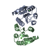
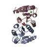
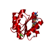
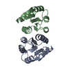
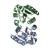
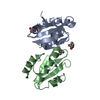
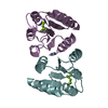
| Title | BeF3 Activated DrrD Receiver Domain | ||||||
 Components Components | DNA BINDING RESPONSE REGULATOR D | ||||||
 Keywords Keywords | DNA BINDING PROTEIN / CheY-like fold / alpha/beta | ||||||
| Function / homology |  Function and homology information Function and homology informationphosphorelay response regulator activity / protein-DNA complex / transcription cis-regulatory region binding / regulation of DNA-templated transcription / DNA-templated transcription / metal ion binding / cytosol Similarity search - Function | ||||||
| Biological species |   Thermotoga maritima (bacteria) Thermotoga maritima (bacteria) | ||||||
| Method |  X-RAY DIFFRACTION / SYNCHROTRON / MOLECULAR REPLACEMENT / Resolution: 2.2 Å X-RAY DIFFRACTION / SYNCHROTRON / MOLECULAR REPLACEMENT / Resolution: 2.2 Å | ||||||
 Authors Authors | Robinson, V.L. / Stock, A.M. | ||||||
 Citation Citation | Journal: J.Biol.Chem. / Year: 2010 Title: Regulation of response regulator autophosphorylation through interdomain contacts. Authors: Barbieri, C.M. / Mack, T.R. / Robinson, V.L. / Miller, M.T. / Stock, A.M. | ||||||
| History |
|
- Structure visualization
| Structure viewer | Molecule: MolmilJmol/JSmol |
|---|
- Downloads & links
-Download
| PDBx/mmCIF format | 3nnn.cif.gz | 63.2 KB | Display | PDBx/mmCIF format |
|---|---|---|---|---|
| PDB format | pdb3nnn.ent.gz | 46.3 KB | Display | PDB format |
| PDBx/mmJSON format | 3nnn.json.gz | Tree view | PDBx/mmJSON format | |
| Others |  Other downloads Other downloads |
-Validation report
| Arichive directory | https://data.pdbj.org/pub/pdb/validation_reports/nn/3nnnftp://data.pdbj.org/pub/pdb/validation_reports/nn/3nnn | HTTPS FTP |
|---|
-Related structure data
| Related structure data |  3nhzC  3nnsC  1kgsS S: Starting model for refinement C: citing same article ( |
|---|---|
| Similar structure data |

-Links
 PDBj
PDBj


- Assembly
| Deposited unit | 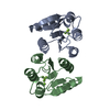
| ||||||||
|---|---|---|---|---|---|---|---|---|---|
| 1 |
| ||||||||
| Unit cell |
|
-Components
| #1: Protein | Mass: 13992.295 Da / Num. of mol.: 2 / Fragment: N-terminal Domain (UNP residues 1-122) Source method: isolated from a genetically manipulated source Source: (gene. exp.) Thermotoga maritima (bacteria) / Gene: DRRD, TM_0399 / Plasmid: pET21b / Production host: #2: Chemical |   Mass: 24.305 Da / Num. of mol.: 2 / Source method: obtained synthetically / Formula: Mg Mass: 24.305 Da / Num. of mol.: 2 / Source method: obtained synthetically / Formula: Mg#3: Chemical |   Mass: 66.007 Da / Num. of mol.: 2 / Source method: obtained synthetically / Formula: BeF3 Mass: 66.007 Da / Num. of mol.: 2 / Source method: obtained synthetically / Formula: BeF3#4: Water | ChemComp-HOH / |  Mass: 18.015 Da / Num. of mol.: 138 / Source method: isolated from a natural source / Formula: H2O Mass: 18.015 Da / Num. of mol.: 138 / Source method: isolated from a natural source / Formula: H2O |
|---|
-Experimental details
-Experiment
| Experiment | Method: X-RAY DIFFRACTION / Number of used crystals: 1 |
|---|
- Sample preparation
| Crystal | Density Matthews: 2.48 Å3/Da / Density % sol: 50.33 % |
|---|---|
| Crystal grow | Temperature: 298 K / Method: vapor diffusion, hanging drop / pH: 4.7 Details: 9-12 % PEG 3350, 0.2M Sodium phosphate monobasic, 6 mM beryllium chloride, 50 mM sodium fluoride, 10 mM magnesium chloride, pH 4.7, VAPOR DIFFUSION, HANGING DROP, temperature 298K |
-Data collection
| Diffraction | Mean temperature: 100 K |
|---|---|
| Diffraction source | Source: SYNCHROTRON / Site: NSLS  / Beamline: X4A / Wavelength: 0.9789 Å / Beamline: X4A / Wavelength: 0.9789 Å |
| Detector | Type: ADSC QUANTUM 4 / Detector: CCD / Date: Jul 31, 2004 |
| Radiation | Monochromator: KOHZU double crystal monochromator with a sagitally focused second crystal. Crystal type Si(III) Protocol: SINGLE WAVELENGTH / Monochromatic (M) / Laue (L): M / Scattering type: x-ray |
| Radiation wavelength | Wavelength: 0.9789 Å / Relative weight: 1 |
| Reflection | Resolution: 2.2→30 Å / Num. all: 14886 / Num. obs: 14831 / % possible obs: 99.7 % / Observed criterion σ(F): 0 / Observed criterion σ(I): -3 / Redundancy: 5.9 % / Rmerge(I) obs: 0.087 |
| Reflection shell | Resolution: 2.2→2.28 Å / Redundancy: 4.3 % / Rmerge(I) obs: 0.41 / Mean I/σ(I) obs: 4.7 / Num. unique all: 1409 / Rsym value: 0.402 / % possible all: 97.8 |
- Processing
| Software |
| ||||||||||||||||||||
|---|---|---|---|---|---|---|---|---|---|---|---|---|---|---|---|---|---|---|---|---|---|
| Refinement | Method to determine structure: MOLECULAR REPLACEMENT Starting model: PDB ENTRY 1KGS Resolution: 2.2→30 Å / σ(F): 0 / Stereochemistry target values: Engh & Huber
| ||||||||||||||||||||
| Refinement step | Cycle: LAST / Resolution: 2.2→30 Å
| ||||||||||||||||||||
| Refine LS restraints |
|
