 Movie
Movie Controller

[English] 日本語
 Yorodumi
- PDB-3nkl: Crystal Structure of UDP-D-Quinovosamine 4-Dehydrogenase from Vib... -
+ Open data

- Basic information
| Entry | Database: PDB / ID: 3nkl | ||||||
|---|---|---|---|---|---|---|---|
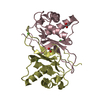
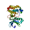
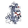
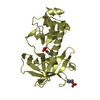
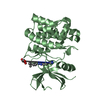
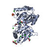
| Title | Crystal Structure of UDP-D-Quinovosamine 4-Dehydrogenase from Vibrio fischeri | ||||||
 Components Components | UDP-D-quinovosamine 4-dehydrogenase | ||||||
 Keywords Keywords | oxidoreductase/lyase / alpha-beta fold / Structural Genomics / PSI-2 / Protein Structure Initiative / Midwest Center for Structural Genomics / MCSG / oxidoreductase-lyase complex | ||||||
| Function / homology |  Function and homology information Function and homology informationLyases; Carbon-oxygen lyases; Hydro-lyases / Oxidoreductases; Acting on the CH-OH group of donors; With NAD+ or NADP+ as acceptor / lyase activity / oxidoreductase activity Similarity search - Function | ||||||
| Biological species |  Vibrio fischeri (bacteria) Vibrio fischeri (bacteria) | ||||||
| Method |  X-RAY DIFFRACTION / SYNCHROTRON / SAD / Resolution: 1.9 Å X-RAY DIFFRACTION / SYNCHROTRON / SAD / Resolution: 1.9 Å | ||||||
 Authors Authors | Kim, Y. / Mack, J. / Buck, K. / Joachimiak, A. / Midwest Center for Structural Genomics (MCSG) | ||||||
 Citation Citation | Journal: To be Published / Year: 2010 Title: Crystal Structure of UDP-D-Quinovosamine 4-Dehydrogenase from Vibrio fischeri Authors: Kim, Y. / Mack, J. / Buck, K. / Joachimiak, A. | ||||||
| History |
|
- Structure visualization
| Structure viewer | Molecule: MolmilJmol/JSmol |
|---|
- Downloads & links
-Download
| PDBx/mmCIF format | 3nkl.cif.gz | 121.7 KB | Display | PDBx/mmCIF format |
|---|---|---|---|---|
| PDB format | pdb3nkl.ent.gz | 94.5 KB | Display | PDB format |
| PDBx/mmJSON format | 3nkl.json.gz | Tree view | PDBx/mmJSON format | |
| Others |  Other downloads Other downloads |
-Validation report
| Arichive directory | https://data.pdbj.org/pub/pdb/validation_reports/nk/3nklftp://data.pdbj.org/pub/pdb/validation_reports/nk/3nkl | HTTPS FTP |
|---|
-Related structure data
| Similar structure data | |
|---|---|
| Other databases |


-Links
 PDBj
PDBj

- Assembly
| Deposited unit | 
| ||||||||
|---|---|---|---|---|---|---|---|---|---|
| 1 |
| ||||||||
| Unit cell |
|
-Components
-Protein , 1 types, 2 molecules AB
| #1: Protein | Mass: 15762.071 Da / Num. of mol.: 2 / Fragment: residues 144-281 Source method: isolated from a genetically manipulated source Source: (gene. exp.) Vibrio fischeri (bacteria) / Strain: ES114 / Gene: VF_0197 / Plasmid: pMCSG7 / Production host: References: UniProt: Q5E8F4, Oxidoreductases; Acting on the CH-OH group of donors; With NAD+ or NADP+ as acceptor, Lyases; Carbon-oxygen lyases; Hydro-lyases |
|---|
-Non-polymers , 5 types, 253 molecules 


| #2: Chemical | ChemComp-SO4 /  Mass: 96.063 Da / Num. of mol.: 5 / Source method: obtained synthetically / Formula: SO4 Mass: 96.063 Da / Num. of mol.: 5 / Source method: obtained synthetically / Formula: SO4#3: Chemical |  Mass: 92.094 Da / Num. of mol.: 2 / Source method: obtained synthetically / Formula: C3H8O3 Mass: 92.094 Da / Num. of mol.: 2 / Source method: obtained synthetically / Formula: C3H8O3#4: Chemical | ChemComp-ACY / |  Mass: 60.052 Da / Num. of mol.: 1 / Source method: obtained synthetically / Formula: C2H4O2 Mass: 60.052 Da / Num. of mol.: 1 / Source method: obtained synthetically / Formula: C2H4O2#5: Chemical | ChemComp-CL / |  Mass: 35.453 Da / Num. of mol.: 1 / Source method: obtained synthetically / Formula: Cl Mass: 35.453 Da / Num. of mol.: 1 / Source method: obtained synthetically / Formula: Cl#6: Water | ChemComp-HOH / | Mass: 18.015 Da / Num. of mol.: 244 / Source method: isolated from a natural source / Formula: H2O |
|---|
-Details
| Has protein modification | Y |
|---|
-Experimental details
-Experiment
| Experiment | Method: X-RAY DIFFRACTION / Number of used crystals: 1 |
|---|
- Sample preparation
| Crystal | Density Matthews: 2.72 Å3/Da / Density % sol: 54.76 % |
|---|---|
| Crystal grow | Temperature: 277 K / Method: vapor diffusion, sitting drop / pH: 7.5 Details: 0.2 M Ammonium sulfate, 0.1 M HEPES pH 7.5, 25 %w/v Polyehtlyene glycol 3,350, VAPOR DIFFUSION, SITTING DROP, temperature 277K |
-Data collection
| Diffraction | Mean temperature: 100 K |
|---|---|
| Diffraction source | Source: SYNCHROTRON / Site: APS  / Beamline: 19-ID / Wavelength: 0.97931 Å / Beamline: 19-ID / Wavelength: 0.97931 Å |
| Detector | Type: ADSC QUANTUM 315r / Detector: CCD / Date: Jun 13, 2010 / Details: mirrors |
| Radiation | Monochromator: double crystal monochromator / Protocol: SINGLE WAVELENGTH / Monochromatic (M) / Laue (L): M / Scattering type: x-ray |
| Radiation wavelength | Wavelength: 0.97931 Å / Relative weight: 1 |
| Reflection | Resolution: 1.9→50 Å / Num. all: 25582 / Num. obs: 25582 / % possible obs: 94.6 % / Observed criterion σ(F): 0 / Observed criterion σ(I): 0 / Redundancy: 4.1 % / Biso Wilson estimate: 24.67 Å2 / Rsym value: 0.051 / Net I/σ(I): 13.3 |
| Reflection shell | Resolution: 1.9→1.93 Å / Redundancy: 3.4 % / Mean I/σ(I) obs: 4.26 / Num. unique all: 832 / Rsym value: 0.341 / % possible all: 62.5 |
- Processing
| Software |
| |||||||||||||||||||||||||||||||||||||||||||||||||||||||||||||||||||||||||||
|---|---|---|---|---|---|---|---|---|---|---|---|---|---|---|---|---|---|---|---|---|---|---|---|---|---|---|---|---|---|---|---|---|---|---|---|---|---|---|---|---|---|---|---|---|---|---|---|---|---|---|---|---|---|---|---|---|---|---|---|---|---|---|---|---|---|---|---|---|---|---|---|---|---|---|---|---|
| Refinement | Method to determine structure: SAD / Resolution: 1.9→42.217 Å / SU ML: 0.24 / Isotropic thermal model: mixed / Cross valid method: THROUGHOUT / σ(F): 0 / Stereochemistry target values: MLHL
| |||||||||||||||||||||||||||||||||||||||||||||||||||||||||||||||||||||||||||
| Solvent computation | Shrinkage radii: 0.9 Å / VDW probe radii: 1.11 Å / Solvent model: FLAT BULK SOLVENT MODEL / Bsol: 42.645 Å2 / ksol: 0.358 e/Å3 | |||||||||||||||||||||||||||||||||||||||||||||||||||||||||||||||||||||||||||
| Displacement parameters | Biso mean: 37 Å2
| |||||||||||||||||||||||||||||||||||||||||||||||||||||||||||||||||||||||||||
| Refinement step | Cycle: LAST / Resolution: 1.9→42.217 Å
| |||||||||||||||||||||||||||||||||||||||||||||||||||||||||||||||||||||||||||
| Refine LS restraints |
| |||||||||||||||||||||||||||||||||||||||||||||||||||||||||||||||||||||||||||
| LS refinement shell | Refine-ID: X-RAY DIFFRACTION
| |||||||||||||||||||||||||||||||||||||||||||||||||||||||||||||||||||||||||||
| Refinement TLS params. | Method: refined / Refine-ID: X-RAY DIFFRACTION
| |||||||||||||||||||||||||||||||||||||||||||||||||||||||||||||||||||||||||||
| Refinement TLS group |
|
