 Movie
Movie Controller

[English] 日本語
 Yorodumi
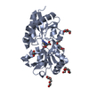
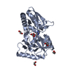
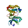
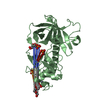
- PDB-5ot8: Structure of the periplasmic binding protein (PBP) NocT-G97S muta... -
+ Open data

- Basic information
| Entry | Database: PDB / ID: 5ot8 | ||||||
|---|---|---|---|---|---|---|---|
| Title | Structure of the periplasmic binding protein (PBP) NocT-G97S mutant from A. tumefaciens C58 in complex with octopine. | ||||||
 Components Components | Nopaline-binding periplasmic protein | ||||||
 Keywords Keywords | PROTEIN BINDING / Agrobacterium tumefaciens / Arginine / Bacterial Proteins / DNA / Bacterial / Gene Expression Regulation / Genes / Ligands / Plant Tumors / Plasmids | ||||||
| Function / homology |  Function and homology information Function and homology informationligand-gated monoatomic ion channel activity / outer membrane-bounded periplasmic space / membrane Similarity search - Function | ||||||
| Biological species |  Agrobacterium fabrum str. C58 (bacteria) Agrobacterium fabrum str. C58 (bacteria) | ||||||
| Method |  X-RAY DIFFRACTION / SYNCHROTRON / MOLECULAR REPLACEMENT / Resolution: 2.35 Å X-RAY DIFFRACTION / SYNCHROTRON / MOLECULAR REPLACEMENT / Resolution: 2.35 Å | ||||||
 Authors Authors | Vigouroux, A. / Morera, S. | ||||||
 Citation Citation | Journal: Sci Rep / Year: 2017 Title: Structural basis for high specificity of octopine binding in the plant pathogen Agrobacterium tumefaciens. Authors: Vigouroux, A. / El Sahili, A. / Lang, J. / Aumont-Nicaise, M. / Dessaux, Y. / Faure, D. / Morera, S. | ||||||
| History |
|
- Structure visualization
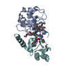
| Structure viewer | Molecule: MolmilJmol/JSmol |
|---|
- Downloads & links
-Download
| PDBx/mmCIF format | 5ot8.cif.gz | 211.2 KB | Display | PDBx/mmCIF format |
|---|---|---|---|---|
| PDB format | pdb5ot8.ent.gz | 168.7 KB | Display | PDB format |
| PDBx/mmJSON format | 5ot8.json.gz | Tree view | PDBx/mmJSON format | |
| Others |  Other downloads Other downloads |
-Validation report
| Arichive directory | https://data.pdbj.org/pub/pdb/validation_reports/ot/5ot8ftp://data.pdbj.org/pub/pdb/validation_reports/ot/5ot8 | HTTPS FTP |
|---|
-Related structure data
| Related structure data | 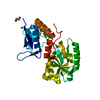 5oreC 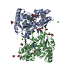 5orgC  5ot9C 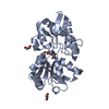 5otaC 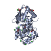 5otcC 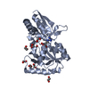 5itpS S: Starting model for refinement C: citing same article ( |
|---|---|
| Similar structure data |


-Links
 PDBj
PDBj


- Assembly
| Deposited unit | 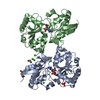
| ||||||||
|---|---|---|---|---|---|---|---|---|---|
| 1 | 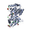
| ||||||||
| 2 | 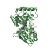
| ||||||||
| Unit cell |
|
-Components
-Protein , 1 types, 2 molecules AB
| #1: Protein | Mass: 29289.650 Da / Num. of mol.: 2 Source method: isolated from a genetically manipulated source Details: HHHHHH : Tag Source: (gene. exp.) Agrobacterium fabrum str. C58 (bacteria)Gene: nocT, Atu6027, AGR_pTi_67 / Production host: |
|---|
-Non-polymers , 6 types, 67 molecules 


| #2: Chemical |  Mass: 246.264 Da / Num. of mol.: 2 / Source method: obtained synthetically / Formula: C9H18N4O4 Mass: 246.264 Da / Num. of mol.: 2 / Source method: obtained synthetically / Formula: C9H18N4O4#3: Chemical |  Mass: 62.068 Da / Num. of mol.: 3 / Source method: obtained synthetically / Formula: C2H6O2 Mass: 62.068 Da / Num. of mol.: 3 / Source method: obtained synthetically / Formula: C2H6O2#4: Chemical | ChemComp-SO4 / |  Mass: 96.063 Da / Num. of mol.: 1 / Source method: obtained synthetically / Formula: SO4 Mass: 96.063 Da / Num. of mol.: 1 / Source method: obtained synthetically / Formula: SO4#5: Chemical | ChemComp-CL /  Mass: 35.453 Da / Num. of mol.: 9 / Source method: obtained synthetically / Formula: Cl Mass: 35.453 Da / Num. of mol.: 9 / Source method: obtained synthetically / Formula: Cl#6: Chemical | ChemComp-PEG / |  Mass: 106.120 Da / Num. of mol.: 1 / Source method: obtained synthetically / Formula: C4H10O3 Mass: 106.120 Da / Num. of mol.: 1 / Source method: obtained synthetically / Formula: C4H10O3#7: Water | ChemComp-HOH / | Mass: 18.015 Da / Num. of mol.: 51 / Source method: isolated from a natural source / Formula: H2O |
|---|
-Experimental details
-Experiment
| Experiment | Method: X-RAY DIFFRACTION / Number of used crystals: 1 |
|---|
- Sample preparation
| Crystal | Density Matthews: 2.51 Å3/Da / Density % sol: 50.99 % |
|---|---|
| Crystal grow | Temperature: 292 K / Method: vapor diffusion, hanging drop / Details: 30% PEG 4K, 0.1 M Tris pH8, 0.1 M LiSO4 |
-Data collection
| Diffraction | Mean temperature: 100 K |
|---|---|
| Diffraction source | Source: SYNCHROTRON / Site: SOLEIL  / Beamline: PROXIMA 1 / Wavelength: 1.006 Å / Beamline: PROXIMA 1 / Wavelength: 1.006 Å |
| Detector | Type: DECTRIS PILATUS 6M / Detector: PIXEL / Date: Mar 17, 2013 |
| Radiation | Protocol: SINGLE WAVELENGTH / Monochromatic (M) / Laue (L): M / Scattering type: x-ray |
| Radiation wavelength | Wavelength: 1.006 Å / Relative weight: 1 |
| Reflection | Resolution: 2.35→50 Å / Num. obs: 23735 / % possible obs: 99.8 % / Redundancy: 10.5 % / Biso Wilson estimate: 57.87 Å2 / Rsym value: 0.093 / Net I/σ(I): 11.6 |
| Reflection shell | Resolution: 2.35→2.49 Å / Mean I/σ(I) obs: 1.8 / Num. unique all: 3814 / Rsym value: 0.715 / % possible all: 99.7 |
- Processing
| Software |
| ||||||||||||||||||||||||||||||||||||||||||||||||||||||||||||||||||||||||||||||||||||||||||||||||||||||||||||||||||
|---|---|---|---|---|---|---|---|---|---|---|---|---|---|---|---|---|---|---|---|---|---|---|---|---|---|---|---|---|---|---|---|---|---|---|---|---|---|---|---|---|---|---|---|---|---|---|---|---|---|---|---|---|---|---|---|---|---|---|---|---|---|---|---|---|---|---|---|---|---|---|---|---|---|---|---|---|---|---|---|---|---|---|---|---|---|---|---|---|---|---|---|---|---|---|---|---|---|---|---|---|---|---|---|---|---|---|---|---|---|---|---|---|---|---|---|
| Refinement | Method to determine structure: MOLECULAR REPLACEMENT Starting model: 5ITP Resolution: 2.35→26.43 Å / Cor.coef. Fo:Fc: 0.954 / Cor.coef. Fo:Fc free: 0.948 / Rfactor Rfree error: 0.001 / SU R Cruickshank DPI: 0.314 / Cross valid method: THROUGHOUT / σ(F): 0 / SU R Blow DPI: 0.284 / SU Rfree Blow DPI: 0.19 / SU Rfree Cruickshank DPI: 0.198
| ||||||||||||||||||||||||||||||||||||||||||||||||||||||||||||||||||||||||||||||||||||||||||||||||||||||||||||||||||
| Displacement parameters | Biso mean: 52.17 Å2
| ||||||||||||||||||||||||||||||||||||||||||||||||||||||||||||||||||||||||||||||||||||||||||||||||||||||||||||||||||
| Refine analyze | Luzzati coordinate error obs: 0.29 Å | ||||||||||||||||||||||||||||||||||||||||||||||||||||||||||||||||||||||||||||||||||||||||||||||||||||||||||||||||||
| Refinement step | Cycle: 1 / Resolution: 2.35→26.43 Å
| ||||||||||||||||||||||||||||||||||||||||||||||||||||||||||||||||||||||||||||||||||||||||||||||||||||||||||||||||||
| Refine LS restraints |
| ||||||||||||||||||||||||||||||||||||||||||||||||||||||||||||||||||||||||||||||||||||||||||||||||||||||||||||||||||
| LS refinement shell | Resolution: 2.35→2.45 Å / Rfactor Rfree error: 0 / Total num. of bins used: 12
| ||||||||||||||||||||||||||||||||||||||||||||||||||||||||||||||||||||||||||||||||||||||||||||||||||||||||||||||||||
| Refinement TLS params. | Method: refined / Refine-ID: X-RAY DIFFRACTION
| ||||||||||||||||||||||||||||||||||||||||||||||||||||||||||||||||||||||||||||||||||||||||||||||||||||||||||||||||||
| Refinement TLS group |
|
