- PDB-3njt: Crystal structure of the R450A mutant of the membrane protein FhaC -
+
Open data
ID or keywords:
Loading...
-
Basic information
Entry
Database: PDB / ID: 3njt
Title
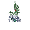
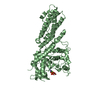
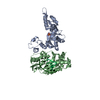
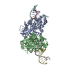
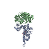
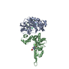
Crystal structure of the R450A mutant of the membrane protein FhaC
Components
Filamentous hemagglutinin transporter protein fhaC
Keywords
PROTEIN TRANSPORT / Membrane protein / beta barrel / Omp85/TpsB transporter family
Function / homology
Function and homology information
type V protein secretion system complex / protein secretion by the type V secretion system / porin activity / pore complex / protein transmembrane transporter activity / monoatomic ion transport / cell outer membrane Similarity search - Function
Two partner secretion pathway transporter / ShlB, POTRA domain / : / POTRA domain / Haemolysin activator HlyB, C-terminal / Haemolysin secretion/activation protein ShlB/FhaC/HecB / Polypeptide-transport-associated, ShlB-type / POTRA domain, ShlB-type / membrane protein fhac: a member of the omp85/tpsb transporter family / membrane protein fhac ...Two partner secretion pathway transporter / ShlB, POTRA domain / : / POTRA domain / Haemolysin activator HlyB, C-terminal / Haemolysin secretion/activation protein ShlB/FhaC/HecB / Polypeptide-transport-associated, ShlB-type / POTRA domain, ShlB-type / membrane protein fhac: a member of the omp85/tpsb transporter family / membrane protein fhac / POTRA domain / POTRA domain profile. / Porin / Ubiquitin-like (UB roll) / Roll / Beta Barrel / Mainly Beta / Alpha Beta Similarity search - Domain/homology
In the structure databanks used in Yorodumi, some data are registered as the other names, "COVID-19 virus" and "2019-nCoV". Here are the details of the virus and the list of structure data.
Jan 31, 2019. EMDB accession codes are about to change! (news from PDBe EMDB page)
EMDB accession codes are about to change! (news from PDBe EMDB page)
The allocation of 4 digits for EMDB accession codes will soon come to an end. Whilst these codes will remain in use, new EMDB accession codes will include an additional digit and will expand incrementally as the available range of codes is exhausted. The current 4-digit format prefixed with “EMD-” (i.e. EMD-XXXX) will advance to a 5-digit format (i.e. EMD-XXXXX), and so on. It is currently estimated that the 4-digit codes will be depleted around Spring 2019, at which point the 5-digit format will come into force.
The EM Navigator/Yorodumi systems omit the EMD- prefix.
Related info.:Q: What is EMD? / ID/Accession-code notation in Yorodumi/EM Navigator
Yorodumi is a browser for structure data from EMDB, PDB, SASBDB, etc.
This page is also the successor to EM Navigator detail page, and also detail information page/front-end page for Omokage search.
The word "yorodu" (or yorozu) is an old Japanese word meaning "ten thousand". "mi" (miru) is to see.
Related info.:EMDB / PDB / SASBDB / Comparison of 3 databanks / Yorodumi Search / Aug 31, 2016. New EM Navigator & Yorodumi / Yorodumi Papers / Jmol/JSmol / Function and homology information / Changes in new EM Navigator and Yorodumi
 Movie
Movie Controller

 Yorodumi
 Open data
 Basic information
 Components
 Keywords
 Function and homology information
 Bordetella pertussis (bacteria)
 X-RAY DIFFRACTION /
  Authors
 Citation
 Structure visualization
 Downloads & links
 Other downloads
 PDBj
PDBj Assembly
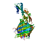
 Sample preparation
 / Beamline: ID14-1 / Wavelength: 0.9334 Å
 Processing