Type: MARMOSAIC 300 mm CCD / Detector: CCD / Date: Oct 12, 2009
Radiation
Monochromator: Si(111) / Protocol: SINGLE WAVELENGTH / Monochromatic (M) / Laue (L): M / Scattering type: x-ray
Radiation wavelength
Wavelength: 1.0781 Å / Relative weight: 1
Reflection
Redundancy: 5.4 % / Av σ(I) over netI: 13.62 / Number: 113045 / Rmerge(I) obs: 0.086 / Χ2: 0.93 / D res high: 2.47 Å / D res low: 50 Å / Num. obs: 21108 / % possible obs: 95.3
Diffraction reflection shell
Highest resolution (Å)
Lowest resolution (Å)
% possible obs (%)
ID
Rmerge(I) obs
Chi squared
Redundancy
6.7
50
87.8
1
0.032
0.783
4.4
5.32
6.7
88.7
1
0.04
0.899
4.7
4.65
5.32
90.6
1
0.036
0.896
4.8
4.22
4.65
92.9
1
0.042
0.879
5
3.92
4.22
93.5
1
0.052
0.912
5.1
3.69
3.92
93
1
0.08
0.73
4.8
3.5
3.69
88.8
1
0.128
0.835
4.4
3.35
3.5
96.5
1
0.099
0.683
5.2
3.22
3.35
97.6
1
0.107
0.947
5.5
3.11
3.22
99.1
1
0.127
0.963
5.7
3.01
3.11
97.6
1
0.136
1.017
5.9
2.93
3.01
99.5
1
0.163
1.04
6
2.85
2.93
97.7
1
0.217
1.01
6.2
2.78
2.85
99.7
1
0.263
1.041
6.2
2.72
2.78
97.7
1
0.332
1.035
6.2
2.66
2.72
99.4
1
0.395
0.915
6
2.61
2.66
99.5
1
0.466
0.852
5.8
2.56
2.61
98.5
1
0.527
1.03
5.5
2.51
2.56
99.3
1
0.566
0.974
5.2
2.47
2.51
93.1
1
0.509
0.97
4.4
Reflection
Resolution: 2.47→50 Å / Num. obs: 21108 / % possible obs: 95.3 % / Redundancy: 5.4 % / Rmerge(I) obs: 0.086 / Χ2: 0.929 / Net I/σ(I): 10.3
Reflection shell
Resolution (Å)
Redundancy (%)
Rmerge(I) obs
Num. unique all
Χ2
% possible all
2.47-2.51
4.4
0.509
1018
0.97
93.1
2.51-2.56
5.2
0.566
1016
0.974
99.3
2.56-2.61
5.5
0.527
1073
1.03
98.5
2.61-2.66
5.8
0.466
1029
0.852
99.5
2.66-2.72
6
0.395
1069
0.915
99.4
2.72-2.78
6.2
0.332
1038
1.035
97.7
2.78-2.85
6.2
0.263
1086
1.041
99.7
2.85-2.93
6.2
0.217
1024
1.01
97.7
2.93-3.01
6
0.163
1108
1.04
99.5
3.01-3.11
5.9
0.136
1026
1.017
97.6
3.11-3.22
5.7
0.127
1061
0.963
99.1
3.22-3.35
5.5
0.107
1100
0.947
97.6
3.35-3.5
5.2
0.099
1036
0.683
96.5
3.5-3.69
4.4
0.128
970
0.835
88.8
3.69-3.92
4.8
0.08
1027
0.73
93
3.92-4.22
5.1
0.052
1050
0.912
93.5
4.22-4.65
5
0.042
1065
0.879
92.9
4.65-5.32
4.8
0.036
1046
0.896
90.6
5.32-6.7
4.7
0.04
1069
0.899
88.7
6.7-50
4.4
0.032
1197
0.783
87.8
-
Phasing
Phasing
Method: molecular replacement
-
Processing
Software
Name
Version
Classification
NB
DENZO
datareduction
SCALEPACK
datascaling
PHENIX
1.5_2
refinement
PDB_EXTRACT
3.1
dataextraction
HKL-2000
datacollection
HKL-2000
datareduction
HKL-2000
datascaling
PHENIX
phasing
Refinement
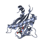
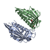
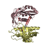
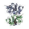
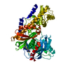
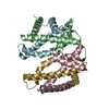
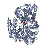
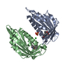
Method to determine structure: MOLECULAR REPLACEMENT Starting model: PDB entry 3JRS, subunit A only
In the structure databanks used in Yorodumi, some data are registered as the other names, "COVID-19 virus" and "2019-nCoV". Here are the details of the virus and the list of structure data.
Jan 31, 2019. EMDB accession codes are about to change! (news from PDBe EMDB page)
EMDB accession codes are about to change! (news from PDBe EMDB page)
The allocation of 4 digits for EMDB accession codes will soon come to an end. Whilst these codes will remain in use, new EMDB accession codes will include an additional digit and will expand incrementally as the available range of codes is exhausted. The current 4-digit format prefixed with “EMD-” (i.e. EMD-XXXX) will advance to a 5-digit format (i.e. EMD-XXXXX), and so on. It is currently estimated that the 4-digit codes will be depleted around Spring 2019, at which point the 5-digit format will come into force.
The EM Navigator/Yorodumi systems omit the EMD- prefix.
Related info.:Q: What is EMD? / ID/Accession-code notation in Yorodumi/EM Navigator
Yorodumi is a browser for structure data from EMDB, PDB, SASBDB, etc.
This page is also the successor to EM Navigator detail page, and also detail information page/front-end page for Omokage search.
The word "yorodu" (or yorozu) is an old Japanese word meaning "ten thousand". "mi" (miru) is to see.
Related info.:EMDB / PDB / SASBDB / Comparison of 3 databanks / Yorodumi Search / Aug 31, 2016. New EM Navigator & Yorodumi / Yorodumi Papers / Jmol/JSmol / Function and homology information / Changes in new EM Navigator and Yorodumi
 Movie
Movie Controller


 Open data
 Basic information
 Components
 Keywords
 Function and homology information
 X-RAY DIFFRACTION /
  Authors
 Citation
 Structure visualization
 Downloads & links
 Other downloads


 PDBj
PDBj Assembly
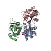


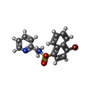


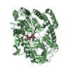
 Mass: 377.256 Da / Num. of mol.: 2 / Source method: obtained synthetically / Formula: C16H13BrN2O2S
 Mass: 96.063 Da / Num. of mol.: 2 / Source method: obtained synthetically / Formula: SO4
 Mass: 22.990 Da / Num. of mol.: 1 / Source method: obtained synthetically / Formula: Na
 Mass: 298.360 Da / Num. of mol.: 1 / Source method: obtained synthetically / Formula: C16H14N2O2S
 Mass: 35.453 Da / Num. of mol.: 1 / Source method: obtained synthetically / Formula: Cl
 Sample preparation
 / Beamline: 21-ID-D / Wavelength: 1.0781 Å
 Processing