 Movie
Movie Controller

[English] 日本語
 Yorodumi
- PDB-3ngr: Crystal structure of Leishmania nucleoside diphosphate kinase b w... -
+ Open data

- Basic information
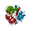
| Entry | Database: PDB / ID: 3ngr | ||||||
|---|---|---|---|---|---|---|---|
| Title | Crystal structure of Leishmania nucleoside diphosphate kinase b with unordered nucleotide-binding loop. | ||||||
 Components Components | Nucleoside diphosphate kinase | ||||||
 Keywords Keywords | TRANSFERASE / NDKb / phosphate ion | ||||||
| Function / homology |  Function and homology information Function and homology informationnucleoside-diphosphate kinase / UTP biosynthetic process / ciliary plasm / CTP biosynthetic process / nucleoside diphosphate kinase activity / GTP biosynthetic process / ATP binding / cytoplasm Similarity search - Function | ||||||
| Biological species |  Leishmania major (eukaryote) Leishmania major (eukaryote) | ||||||
| Method |  X-RAY DIFFRACTION / SYNCHROTRON / MOLECULAR REPLACEMENT / Resolution: 2.95 Å X-RAY DIFFRACTION / SYNCHROTRON / MOLECULAR REPLACEMENT / Resolution: 2.95 Å | ||||||
 Authors Authors | Trindade, D.M. / Sousa, T.A.C.B. / Tonoli, C.C.C. / Santos, C.R. / Arni, R.K. / Ward, R.J. / Oliveira, A.H.C. / Murakami, M.T. | ||||||
 Citation Citation | Journal: Mol Biosyst / Year: 2011 Title: Molecular adaptability of nucleoside diphosphate kinase b from trypanosomatid parasites: stability, oligomerization and structural determinants of nucleotide binding. Authors: Souza, T.A. / Trindade, D.M. / Tonoli, C.C. / Santos, C.R. / Ward, R.J. / Arni, R.K. / Oliveira, A.H. / Murakami, M.T. | ||||||
| History |
|
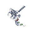
- Structure visualization
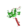
| Structure viewer | Molecule: MolmilJmol/JSmol |
|---|
- Downloads & links
-Download
| PDBx/mmCIF format | 3ngr.cif.gz | 37.3 KB | Display | PDBx/mmCIF format |
|---|---|---|---|---|
| PDB format | pdb3ngr.ent.gz | 25.1 KB | Display | PDB format |
| PDBx/mmJSON format | 3ngr.json.gz | Tree view | PDBx/mmJSON format | |
| Others |  Other downloads Other downloads |
-Validation report
| Arichive directory | https://data.pdbj.org/pub/pdb/validation_reports/ng/3ngrftp://data.pdbj.org/pub/pdb/validation_reports/ng/3ngr | HTTPS FTP |
|---|
-Related structure data
| Related structure data | 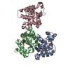 3ngsC 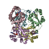 3ngtC 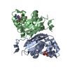 3nguC 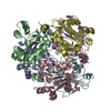 3prvC 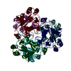 1nskS C: citing same article ( S: Starting model for refinement |
|---|---|
| Similar structure data |


-Links
 PDBj
PDBj- Assembly
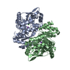
| Deposited unit | 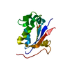
| ||||||||
|---|---|---|---|---|---|---|---|---|---|
| 1 | x 6
| ||||||||
| Unit cell |
|
-Components
| #1: Protein | Mass: 16660.953 Da / Num. of mol.: 1 Source method: isolated from a genetically manipulated source Source: (gene. exp.) Leishmania major (eukaryote) / Gene: L1648.07, LmjF32.2950, NDKb / Plasmid: pET28a / Production host:  |
|---|---|
| #2: Chemical | ChemComp-PO4 /   Mass: 94.971 Da / Num. of mol.: 1 / Source method: obtained synthetically / Formula: PO4 Mass: 94.971 Da / Num. of mol.: 1 / Source method: obtained synthetically / Formula: PO4 |
| #3: Water | ChemComp-HOH /  Mass: 18.015 Da / Num. of mol.: 17 / Source method: isolated from a natural source / Formula: H2O Mass: 18.015 Da / Num. of mol.: 17 / Source method: isolated from a natural source / Formula: H2O |
-Experimental details
-Experiment
| Experiment | Method: X-RAY DIFFRACTION / Number of used crystals: 1 |
|---|
- Sample preparation
| Crystal | Density Matthews: 4.1 Å3/Da / Density % sol: 70 % |
|---|---|
| Crystal grow | Temperature: 291 K / Method: vapor diffusion, sitting drop Details: sodium/potassium phosphate, pH pH 4.5, VAPOR DIFFUSION, SITTING DROP, temperature 291K PH range: pH 4.5 |
-Data collection
| Diffraction | Mean temperature: 100 K |
|---|---|
| Diffraction source | Source: SYNCHROTRON / Site: LNLS  / Beamline: W01B-MX2 / Wavelength: 1.458 Å / Beamline: W01B-MX2 / Wavelength: 1.458 Å |
| Detector | Type: MARMOSAIC 225 mm CCD / Detector: CCD / Date: Mar 10, 2010 |
| Radiation | Monochromator: silicon double-crystal / Protocol: SINGLE WAVELENGTH / Monochromatic (M) / Laue (L): M / Scattering type: x-ray |
| Radiation wavelength | Wavelength: 1.458 Å / Relative weight: 1 |
| Reflection | Resolution: 2.95→104.59 Å / Num. obs: 6148 / % possible obs: 98 % / Redundancy: 11.2 % / Rmerge(I) obs: 0.086 |
| Reflection shell | Resolution: 2.95→3.06 Å / Redundancy: 7 % / Rmerge(I) obs: 0.347 / Mean I/σ(I) obs: 4.5 / % possible all: 86.5 |
- Processing
| Software |
| ||||||||||||||||||||||||||||||||||||||||||||||||||||||||||||||||||||||||||||||||||||||||||||||||||||||||||||||||||||||||||||||||||||||||||||||||||||||||||||||||||||||||||
|---|---|---|---|---|---|---|---|---|---|---|---|---|---|---|---|---|---|---|---|---|---|---|---|---|---|---|---|---|---|---|---|---|---|---|---|---|---|---|---|---|---|---|---|---|---|---|---|---|---|---|---|---|---|---|---|---|---|---|---|---|---|---|---|---|---|---|---|---|---|---|---|---|---|---|---|---|---|---|---|---|---|---|---|---|---|---|---|---|---|---|---|---|---|---|---|---|---|---|---|---|---|---|---|---|---|---|---|---|---|---|---|---|---|---|---|---|---|---|---|---|---|---|---|---|---|---|---|---|---|---|---|---|---|---|---|---|---|---|---|---|---|---|---|---|---|---|---|---|---|---|---|---|---|---|---|---|---|---|---|---|---|---|---|---|---|---|---|---|---|---|---|
| Refinement | Method to determine structure: MOLECULAR REPLACEMENT Starting model: PDB entry: 1NSK Resolution: 2.95→104.59 Å / Cor.coef. Fo:Fc: 0.912 / Cor.coef. Fo:Fc free: 0.834 / SU B: 18.685 / SU ML: 0.353 / Cross valid method: THROUGHOUT / ESU R: 0.489 / ESU R Free: 0.417 / Stereochemistry target values: MAXIMUM LIKELIHOOD / Details: HYDROGENS HAVE BEEN ADDED IN THE RIDING POSITIONS
| ||||||||||||||||||||||||||||||||||||||||||||||||||||||||||||||||||||||||||||||||||||||||||||||||||||||||||||||||||||||||||||||||||||||||||||||||||||||||||||||||||||||||||
| Solvent computation | Ion probe radii: 0.8 Å / Shrinkage radii: 0 Å / VDW probe radii: 1.4 Å / Solvent model: BABINET MODEL WITH MASK | ||||||||||||||||||||||||||||||||||||||||||||||||||||||||||||||||||||||||||||||||||||||||||||||||||||||||||||||||||||||||||||||||||||||||||||||||||||||||||||||||||||||||||
| Displacement parameters | Biso mean: 63.55 Å2
| ||||||||||||||||||||||||||||||||||||||||||||||||||||||||||||||||||||||||||||||||||||||||||||||||||||||||||||||||||||||||||||||||||||||||||||||||||||||||||||||||||||||||||
| Refinement step | Cycle: LAST / Resolution: 2.95→104.59 Å
| ||||||||||||||||||||||||||||||||||||||||||||||||||||||||||||||||||||||||||||||||||||||||||||||||||||||||||||||||||||||||||||||||||||||||||||||||||||||||||||||||||||||||||
| Refine LS restraints |
| ||||||||||||||||||||||||||||||||||||||||||||||||||||||||||||||||||||||||||||||||||||||||||||||||||||||||||||||||||||||||||||||||||||||||||||||||||||||||||||||||||||||||||
| LS refinement shell | Resolution: 2.95→3.025 Å / Total num. of bins used: 20
|
