Shrinkage radii: 0.9 Å / VDW probe radii: 1 Å Solvent model: FLAT BULK SOLVENT MODEL HYDROGENS ADDED IN THE RIDING POSITION DURING REFINEMENT Bsol: 67.273 Å2 / ksol: 0.397 e/Å3
Displacement parameters
Baniso -1
Baniso -2
Baniso -3
1-
3.3884 Å2
-0 Å2
0 Å2
2-
-
-5.3636 Å2
-0 Å2
3-
-
-
1.9753 Å2
Refinement step
Cycle: LAST / Resolution: 1.551→33.036 Å
Protein
Nucleic acid
Ligand
Solvent
Total
Num. atoms
2263
0
0
340
2603
Refine LS restraints
Refine-ID
Type
Dev ideal
Number
X-RAY DIFFRACTION
f_bond_d
0.013
2408
X-RAY DIFFRACTION
f_angle_d
1.333
3296
X-RAY DIFFRACTION
f_dihedral_angle_d
14.726
909
X-RAY DIFFRACTION
f_chiral_restr
0.082
389
X-RAY DIFFRACTION
f_plane_restr
0.007
442
LS refinement shell
Resolution (Å)
Rfactor Rfree
Num. reflection Rfree
Rfactor Rwork
Num. reflection Rwork
Refine-ID
% reflection obs (%)
1.551-1.6064
0.3035
225
0.2534
4388
X-RAY DIFFRACTION
96
1.6064-1.6708
0.2724
232
0.2279
4514
X-RAY DIFFRACTION
98
1.6708-1.7468
0.2646
229
0.2049
4495
X-RAY DIFFRACTION
98
1.7468-1.8389
0.2268
230
0.1925
4482
X-RAY DIFFRACTION
97
1.8389-1.9541
0.2377
216
0.1953
4510
X-RAY DIFFRACTION
97
1.9541-2.1049
0.2002
226
0.1654
4528
X-RAY DIFFRACTION
98
2.1049-2.3167
0.177
217
0.1513
4579
X-RAY DIFFRACTION
98
2.3167-2.6518
0.1745
225
0.1441
4597
X-RAY DIFFRACTION
98
2.6518-3.3405
0.1611
226
0.1537
4657
X-RAY DIFFRACTION
99
3.3405-33.0438
0.1738
232
0.1622
4796
X-RAY DIFFRACTION
98
+
About Yorodumi
-
News
-
Feb 9, 2022. New format data for meta-information of EMDB entries
New format data for meta-information of EMDB entries
Version 3 of the EMDB header file is now the official format.
The previous official version 1.9 will be removed from the archive.
In the structure databanks used in Yorodumi, some data are registered as the other names, "COVID-19 virus" and "2019-nCoV". Here are the details of the virus and the list of structure data.
Jan 31, 2019. EMDB accession codes are about to change! (news from PDBe EMDB page)
EMDB accession codes are about to change! (news from PDBe EMDB page)
The allocation of 4 digits for EMDB accession codes will soon come to an end. Whilst these codes will remain in use, new EMDB accession codes will include an additional digit and will expand incrementally as the available range of codes is exhausted. The current 4-digit format prefixed with “EMD-” (i.e. EMD-XXXX) will advance to a 5-digit format (i.e. EMD-XXXXX), and so on. It is currently estimated that the 4-digit codes will be depleted around Spring 2019, at which point the 5-digit format will come into force.
The EM Navigator/Yorodumi systems omit the EMD- prefix.
Related info.:Q: What is EMD? / ID/Accession-code notation in Yorodumi/EM Navigator
Yorodumi is a browser for structure data from EMDB, PDB, SASBDB, etc.
This page is also the successor to EM Navigator detail page, and also detail information page/front-end page for Omokage search.
The word "yorodu" (or yorozu) is an old Japanese word meaning "ten thousand". "mi" (miru) is to see.
Related info.:EMDB / PDB / SASBDB / Comparison of 3 databanks / Yorodumi Search / Aug 31, 2016. New EM Navigator & Yorodumi / Yorodumi Papers / Jmol/JSmol / Function and homology information / Changes in new EM Navigator and Yorodumi
 Movie
Movie Controller

 Yorodumi
 Open data
 Basic information
 Components
 Keywords
 Function and homology information
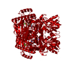
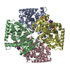
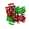
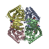
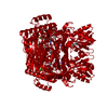
 Salinibacter ruber (bacteria)
 X-RAY DIFFRACTION /
  Authors
 Citation
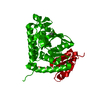
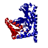
 Structure visualization
 Downloads & links
 Other downloads
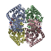

 PDBj
PDBj


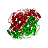
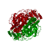
 Assembly
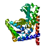
 Mass: 18.015 Da / Num. of mol.: 340 / Source method: isolated from a natural source / Formula: H2O
 Sample preparation
 Processing