[English] 日本語
 Yorodumi
- PDB-4kxu: Human transketolase in covalent complex with donor ketose D-fruct... -
+ Open data
- Basic information
| Entry | Database: PDB / ID: 4kxu | |||||||||
|---|---|---|---|---|---|---|---|---|---|---|
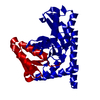
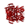
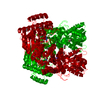
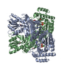
| Title | Human transketolase in covalent complex with donor ketose D-fructose-6-phosphate | |||||||||
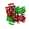
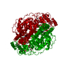
 Components Components | Transketolase | |||||||||
 Keywords Keywords | TRANSFERASE / thiamin diphosphate / enzyme catalysis / pentose phosphate pathway | |||||||||
| Function / homology |  Function and homology information Function and homology informationD-xylulose 5-phosphate biosynthetic process / Insulin effects increased synthesis of Xylulose-5-Phosphate / Pentose phosphate pathway / transketolase / transketolase activity / pentose-phosphate shunt, non-oxidative branch / pentose-phosphate shunt / NFE2L2 regulates pentose phosphate pathway genes / glyceraldehyde-3-phosphate biosynthetic process / regulation of growth ...D-xylulose 5-phosphate biosynthetic process / Insulin effects increased synthesis of Xylulose-5-Phosphate / Pentose phosphate pathway / transketolase / transketolase activity / pentose-phosphate shunt, non-oxidative branch / pentose-phosphate shunt / NFE2L2 regulates pentose phosphate pathway genes / glyceraldehyde-3-phosphate biosynthetic process / regulation of growth / thiamine pyrophosphate binding / peroxisome / vesicle / nuclear body / calcium ion binding / endoplasmic reticulum membrane / magnesium ion binding / protein homodimerization activity / extracellular exosome / nucleoplasm / cytosol Similarity search - Function | |||||||||
| Biological species |  Homo sapiens (human) Homo sapiens (human) | |||||||||
| Method |  X-RAY DIFFRACTION / X-RAY DIFFRACTION /  SYNCHROTRON / SYNCHROTRON /  FOURIER SYNTHESIS / Resolution: 0.98 Å FOURIER SYNTHESIS / Resolution: 0.98 Å | |||||||||
 Authors Authors | Neumann, P. / Luedtke, S. / Ficner, R. / Tittmann, K. | |||||||||
 Citation Citation |  Journal: Nat Chem / Year: 2013 Journal: Nat Chem / Year: 2013Title: Sub-angstrom-resolution crystallography reveals physical distortions that enhance reactivity of a covalent enzymatic intermediate. Authors: Ludtke, S. / Neumann, P. / Erixon, K.M. / Leeper, F. / Kluger, R. / Ficner, R. / Tittmann, K. | |||||||||
| History |
|
- Structure visualization
| Structure viewer | Molecule:  Molmil Molmil Jmol/JSmol Jmol/JSmol |
|---|
- Downloads & links
- Download
| PDBx/mmCIF format |  4kxu.cif.gz 4kxu.cif.gz | 320.2 KB | Display |  PDBx/mmCIF format PDBx/mmCIF format |
|---|---|---|---|---|
| PDB format |  pdb4kxu.ent.gz pdb4kxu.ent.gz | 254.4 KB | Display |  PDB format PDB format |
| PDBx/mmJSON format |  4kxu.json.gz 4kxu.json.gz | Tree view |  PDBx/mmJSON format PDBx/mmJSON format | |
| Others |  Other downloads Other downloads |
-Validation report
| Arichive directory |  https://data.pdbj.org/pub/pdb/validation_reports/kx/4kxu https://data.pdbj.org/pub/pdb/validation_reports/kx/4kxu ftp://data.pdbj.org/pub/pdb/validation_reports/kx/4kxu ftp://data.pdbj.org/pub/pdb/validation_reports/kx/4kxu | HTTPS FTP |
|---|
-Related structure data
| Related structure data | 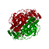 4kxvC  4kxwC  4kxxC  4kxyC 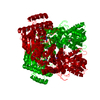 3mosS S: Starting model for refinement C: citing same article ( |
|---|---|
| Similar structure data |
- Links
Links
- Assembly
| Deposited unit | 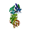
| |||||||||
|---|---|---|---|---|---|---|---|---|---|---|
| 1 | 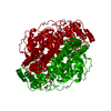
| |||||||||
| Unit cell |
| |||||||||
| Components on special symmetry positions |
|
- Components
-Protein / Sugars , 2 types, 2 molecules A

| #1: Protein | Mass: 69647.461 Da / Num. of mol.: 1 Source method: isolated from a genetically manipulated source Source: (gene. exp.)  Homo sapiens (human) / Gene: TKT / Plasmid: pET-28a / Production host: Homo sapiens (human) / Gene: TKT / Plasmid: pET-28a / Production host:  |
|---|---|
| #5: Sugar | ChemComp-S6P / |
-Non-polymers , 5 types, 834 molecules 


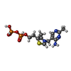


| #2: Chemical | ChemComp-EDO / #3: Chemical | ChemComp-MG / | #4: Chemical | ChemComp-NA / | #6: Chemical | ChemComp-TPP / | #7: Water | ChemComp-HOH / | |
|---|
-Details
| Nonpolymer details | S6P A1014 IS A HYDROLYZED |
|---|
-Experimental details
-Experiment
| Experiment | Method:  X-RAY DIFFRACTION / Number of used crystals: 1 X-RAY DIFFRACTION / Number of used crystals: 1 |
|---|
- Sample preparation
| Crystal | Density Matthews: 2.13 Å3/Da / Density % sol: 42.3 % |
|---|---|
| Crystal grow | Temperature: 279 K / Method: vapor diffusion, hanging drop / pH: 7.9 Details: Reservoir mixture of 13.5-15 % PEG 6000 (w/v), 4% PEG 400 (v/v), 2 % glycerol (v/v) , pH 7.9, VAPOR DIFFUSION, HANGING DROP, temperature 279.0K |
-Data collection
| Diffraction | Mean temperature: 100 K | |||||||||||||||||||||||||||||||||||||||||||||||||||||||||||||||||||||||||||||||||||||||||||
|---|---|---|---|---|---|---|---|---|---|---|---|---|---|---|---|---|---|---|---|---|---|---|---|---|---|---|---|---|---|---|---|---|---|---|---|---|---|---|---|---|---|---|---|---|---|---|---|---|---|---|---|---|---|---|---|---|---|---|---|---|---|---|---|---|---|---|---|---|---|---|---|---|---|---|---|---|---|---|---|---|---|---|---|---|---|---|---|---|---|---|---|---|
| Diffraction source | Source:  SYNCHROTRON / Site: SYNCHROTRON / Site:  ESRF ESRF  / Beamline: ID14-4 / Wavelength: 0.93939 Å / Beamline: ID14-4 / Wavelength: 0.93939 Å | |||||||||||||||||||||||||||||||||||||||||||||||||||||||||||||||||||||||||||||||||||||||||||
| Detector | Type: ADSC QUANTUM 315r / Detector: CCD / Date: Nov 22, 2011 / Details: mirrors | |||||||||||||||||||||||||||||||||||||||||||||||||||||||||||||||||||||||||||||||||||||||||||
| Radiation | Monochromator: GRAPHITE / Protocol: SINGLE WAVELENGTH / Monochromatic (M) / Laue (L): M / Scattering type: x-ray | |||||||||||||||||||||||||||||||||||||||||||||||||||||||||||||||||||||||||||||||||||||||||||
| Radiation wavelength | Wavelength: 0.93939 Å / Relative weight: 1 | |||||||||||||||||||||||||||||||||||||||||||||||||||||||||||||||||||||||||||||||||||||||||||
| Reflection | Resolution: 0.98→30 Å / Num. all: 300587 / Num. obs: 300587 / % possible obs: 91.8 % / Observed criterion σ(F): 0 / Observed criterion σ(I): -3 / Redundancy: 4.5 % / Biso Wilson estimate: 9.844 Å2 / Rmerge(I) obs: 0.051 / Net I/σ(I): 16.09 | |||||||||||||||||||||||||||||||||||||||||||||||||||||||||||||||||||||||||||||||||||||||||||
| Reflection shell |
|
- Processing
| Software |
| |||||||||||||||||||||||||||||||||
|---|---|---|---|---|---|---|---|---|---|---|---|---|---|---|---|---|---|---|---|---|---|---|---|---|---|---|---|---|---|---|---|---|---|---|
| Refinement | Method to determine structure:  FOURIER SYNTHESIS FOURIER SYNTHESISStarting model: PDB ENTRY 3MOS Resolution: 0.98→30 Å / Num. parameters: 57058 / Num. restraintsaints: 62047 / Occupancy max: 1 / Occupancy min: 0.04 / Cross valid method: FREE R / σ(F): 0 / σ(I): 0 / Stereochemistry target values: ENGH AND HUBER
| |||||||||||||||||||||||||||||||||
| Solvent computation | Solvent model: MOEWS & KRETSINGER, J.MOL.BIOL.91(1973)201-228 | |||||||||||||||||||||||||||||||||
| Displacement parameters | Biso max: 184.87 Å2 / Biso mean: 13.6034 Å2 / Biso min: 2.11 Å2 | |||||||||||||||||||||||||||||||||
| Refinement step | Cycle: LAST / Resolution: 0.98→30 Å
| |||||||||||||||||||||||||||||||||
| Refine LS restraints |
|
 Movie
Movie Controller




 PDBj
PDBj


